3RGO

Crystal Structure of PTPMT1


Experimental Data Snapshot

  • Method: X-RAY DIFFRACTION
  • Resolution: 1.93 Å
  • R-Value Free: 0.243 
  • R-Value Work: 0.227 
  • R-Value Observed: 0.228 

wwPDB Validation   3D Report Full Report


This is version 1.5 of the entry. See complete history


Literature

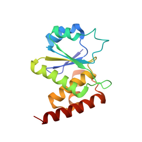
Structural and functional analysis of PTPMT1, a phosphatase required for cardiolipin synthesis.

Xiao, J.Engel, J.L.Zhang, J.Chen, M.J.Manning, G.Dixon, J.E.

(2011) Proc Natl Acad Sci U S A 108: 11860-11865

  • DOI: https://doi.org/10.1073/pnas.1109290108
  • Primary Citation of Related Structures:  
    3RGO, 3RGQ

  • PubMed Abstract: 

    PTPMT1 (PTP localized to the Mitochondrion 1) is a member of the protein tyrosine phosphatase superfamily that is localized exclusively to the mitochondrion. We recently reported that PTPMT1 dephosphorylates phosphatidylglycerol phosphate, an essential intermediate of cardiolipin biosynthesis. To gain further insights into the molecular basis of PTPMT1 function, we determined the crystal structures of the phosphatase domain of PTPMT1. PTPMT1 exhibits a canonical protein tyrosine phosphatase domain fold, resembling many dual-specificity phosphatases such as phosphatase and tensin homolog and vaccinia H1-related phosphatase. We also determined the structure of the catalytically inactive phosphatase in complex with a surrogate substrate, phosphatidylinositol 5-phosphate, which sheds light on the substrate recognition and specificity of PTPMT1. Comparison of the apo and substrate-bound structures of PTPMT1 suggests that it undergoes significant conformational change during catalysis, and we further demonstrated that an evolutionarily conserved EEYE loop is important for its activity.


  • Organizational Affiliation

    Department of Pharmacology, University of California, La Jolla, CA 92093, USA.


Macromolecules
Find similar proteins by:  (by identity cutoff)  |  3D Structure
Entity ID: 1
MoleculeChains Sequence LengthOrganismDetailsImage
Protein-tyrosine phosphatase mitochondrial 1157Mus musculusMutation(s): 0 
Gene Names: PlipPtpmt1
EC: 3.1.3.16 (PDB Primary Data), 3.1.3.48 (PDB Primary Data)
UniProt & NIH Common Fund Data Resources
Find proteins for Q66GT5 (Mus musculus)
Explore Q66GT5 
Go to UniProtKB:  Q66GT5
IMPC:  MGI:1913711
Entity Groups  
Sequence Clusters30% Identity50% Identity70% Identity90% Identity95% Identity100% Identity
UniProt GroupQ66GT5
Sequence Annotations
Expand
  • Reference Sequence
Experimental Data & Validation

Experimental Data

  • Method: X-RAY DIFFRACTION
  • Resolution: 1.93 Å
  • R-Value Free: 0.243 
  • R-Value Work: 0.227 
  • R-Value Observed: 0.228 
  • Space Group: P 43 21 2
Unit Cell:
Length ( Å )Angle ( ˚ )
a = 36.453α = 90
b = 36.453β = 90
c = 230.024γ = 90
Software Package:
Software NamePurpose
DENZOdata reduction
SCALEPACKdata scaling
PHENIXrefinement
PDB_EXTRACTdata extraction
HKL-2000data reduction
HKL-2000data scaling
PHENIXphasing

Structure Validation

View Full Validation Report


Entry History 

Deposition Data

Revision History  (Full details and data files)

  • Version 1.0: 2011-07-06
    Type: Initial release
  • Version 1.1: 2011-07-13
    Changes: Version format compliance
  • Version 1.2: 2011-07-20
    Changes: Database references
  • Version 1.3: 2011-08-03
    Changes: Database references
  • Version 1.4: 2017-11-08
    Changes: Refinement description
  • Version 1.5: 2024-02-21
    Changes: Data collection, Database references, Derived calculations