3RGB

Crystal structure of particulate methane monooxygenase from Methylococcus capsulatus (Bath)


Experimental Data Snapshot

  • Method: X-RAY DIFFRACTION
  • Resolution: 2.80 Å
  • R-Value Free: 0.296 
  • R-Value Work: 0.269 
  • R-Value Observed: 0.270 

wwPDB Validation   3D Report Full Report


This is version 1.2 of the entry. See complete history


Literature

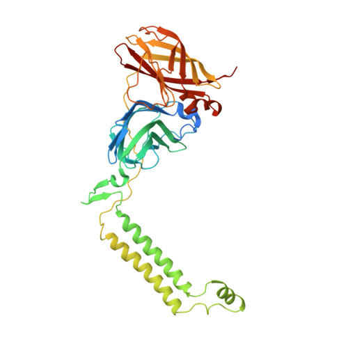
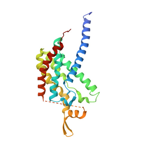
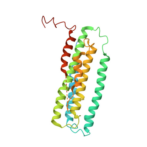
Crystal Structure and Characterization of Particulate Methane Monooxygenase from Methylocystis species Strain M.

Smith, S.M.Rawat, S.Telser, J.Hoffman, B.M.Stemmler, T.L.Rosenzweig, A.C.

(2011) Biochemistry 50: 10231-10240

  • DOI: https://doi.org/10.1021/bi200801z
  • Primary Citation of Related Structures:  
    3RFR, 3RGB

  • PubMed Abstract: 

    Particulate methane monooxygenase (pMMO) is an integral membrane metalloenzyme that oxidizes methane to methanol in methanotrophic bacteria. Previous biochemical and structural studies of pMMO have focused on preparations from Methylococcus capsulatus (Bath) and Methylosinus trichosporium OB3b. A pMMO from a third organism, Methylocystis species strain M, has been isolated and characterized. Both membrane-bound and solubilized Methylocystis sp. strain M pMMO contain ~2 copper ions per 100 kDa protomer and exhibit copper-dependent propylene epoxidation activity. Spectroscopic data indicate that Methylocystis sp. strain M pMMO contains a mixture of Cu(I) and Cu(II), of which the latter exhibits two distinct type 2 Cu(II) electron paramagnetic resonance (EPR) signals. Extended X-ray absorption fine structure (EXAFS) data are best fit with a mixture of Cu-O/N and Cu-Cu ligand environments with a Cu-Cu interaction at 2.52-2.64 Å. The crystal structure of Methylocystis sp. strain M pMMO was determined to 2.68 Å resolution and is the best quality pMMO structure obtained to date. It provides a revised model for the pmoA and pmoC subunits and has led to an improved model of M. capsulatus (Bath) pMMO. In these new structures, the intramembrane zinc/copper binding site has a different coordination environment from that in previous models.


  • Organizational Affiliation

    Departments of Molecular Biosciences and of Chemistry, Northwestern University, Evanston, Illinois 60208, United States.


Macromolecules
Find similar proteins by:  (by identity cutoff)  |  3D Structure
Entity ID: 1
MoleculeChains Sequence LengthOrganismDetailsImage
Methane monooxygenase subunit B2A,
D [auth E],
E [auth I]
414Methylococcus capsulatusMutation(s): 0 
EC: 1.14.13.25
Membrane Entity: Yes 
UniProt
Find proteins for G1UBD1 (Methylococcus capsulatus (strain ATCC 33009 / NCIMB 11132 / Bath))
Explore G1UBD1 
Go to UniProtKB:  G1UBD1
Entity Groups  
Sequence Clusters30% Identity50% Identity70% Identity90% Identity95% Identity100% Identity
UniProt GroupG1UBD1
Sequence Annotations
Expand
  • Reference Sequence
Find similar proteins by:  (by identity cutoff)  |  3D Structure
Entity ID: 2
MoleculeChains Sequence LengthOrganismDetailsImage
Methane monooxygenase subunit A2B,
F [auth J],
G [auth F]
247Methylococcus capsulatusMutation(s): 0 
EC: 1.14.13.25
Membrane Entity: Yes 
UniProt
Find proteins for Q607G3 (Methylococcus capsulatus (strain ATCC 33009 / NCIMB 11132 / Bath))
Explore Q607G3 
Go to UniProtKB:  Q607G3
Entity Groups  
Sequence Clusters30% Identity50% Identity70% Identity90% Identity95% Identity100% Identity
UniProt GroupQ607G3
Sequence Annotations
Expand
  • Reference Sequence
Find similar proteins by:  (by identity cutoff)  |  3D Structure
Entity ID: 3
MoleculeChains Sequence LengthOrganismDetailsImage
Methane monooxygenase subunit C2C,
H [auth K],
I [auth G]
289Methylococcus capsulatusMutation(s): 0 
EC: 1.14.13.25
Membrane Entity: Yes 
UniProt
Find proteins for Q603F1 (Methylococcus capsulatus (strain ATCC 33009 / NCIMB 11132 / Bath))
Explore Q603F1 
Go to UniProtKB:  Q603F1
Entity Groups  
Sequence Clusters30% Identity50% Identity70% Identity90% Identity95% Identity100% Identity
UniProt GroupQ603F1
Sequence Annotations
Expand
  • Reference Sequence
Small Molecules
Ligands 3 Unique
IDChains Name / Formula / InChI Key2D Diagram3D Interactions
CUA
Query on CUA

Download Ideal Coordinates CCD File 
L [auth A],
Q [auth E],
T [auth I]
DINUCLEAR COPPER ION
Cu2
ALKZAGKDWUSJED-UHFFFAOYSA-N
ZN
Query on ZN

Download Ideal Coordinates CCD File 
K [auth A]
M [auth B]
N [auth C]
P [auth E]
S [auth I]
K [auth A],
M [auth B],
N [auth C],
P [auth E],
S [auth I],
U [auth J],
V [auth F],
W [auth K],
X [auth G]
ZINC ION
Zn
PTFCDOFLOPIGGS-UHFFFAOYSA-N
CU
Query on CU

Download Ideal Coordinates CCD File 
J [auth A],
O [auth E],
R [auth I]
COPPER (II) ION
Cu
JPVYNHNXODAKFH-UHFFFAOYSA-N
Experimental Data & Validation

Experimental Data

  • Method: X-RAY DIFFRACTION
  • Resolution: 2.80 Å
  • R-Value Free: 0.296 
  • R-Value Work: 0.269 
  • R-Value Observed: 0.270 
  • Space Group: P 42 21 2
Unit Cell:
Length ( Å )Angle ( ˚ )
a = 264.14α = 90
b = 264.14β = 90
c = 150.01γ = 90
Software Package:
Software NamePurpose
SOLVEphasing
REFMACrefinement
XDSdata reduction
SCALAdata scaling

Structure Validation

View Full Validation Report


Entry History 

Deposition Data

Revision History  (Full details and data files)

  • Version 1.0: 2011-11-23
    Type: Initial release
  • Version 1.1: 2011-12-07
    Changes: Database references
  • Version 1.2: 2024-02-21
    Changes: Data collection, Database references, Derived calculations, Refinement description