3RG9

Trypanosoma brucei dihydrofolate reductase (TbDHFR) in complex with WR99210


Experimental Data Snapshot

  • Method: X-RAY DIFFRACTION
  • Resolution: 2.00 Å
  • R-Value Free: 0.221 
  • R-Value Work: 0.183 
  • R-Value Observed: 0.183 

wwPDB Validation   3D Report Full Report


Ligand Structure Quality Assessment 


This is version 1.2 of the entry. See complete history


Literature

Trypanosomal dihydrofolate reductase reveals natural antifolate resistance

Vanichtanankul, J.Taweechai, S.Yuvaniyama, J.Vilaivan, T.Chitnumsub, P.Kamchonwongpaisan, S.Yuthavong, Y.

(2011) ACS Chem Biol 6: 905-911

  • DOI: https://doi.org/10.1021/cb200124r
  • Primary Citation of Related Structures:  
    3QFX, 3QG2, 3QGT, 3RG9

  • PubMed Abstract: 

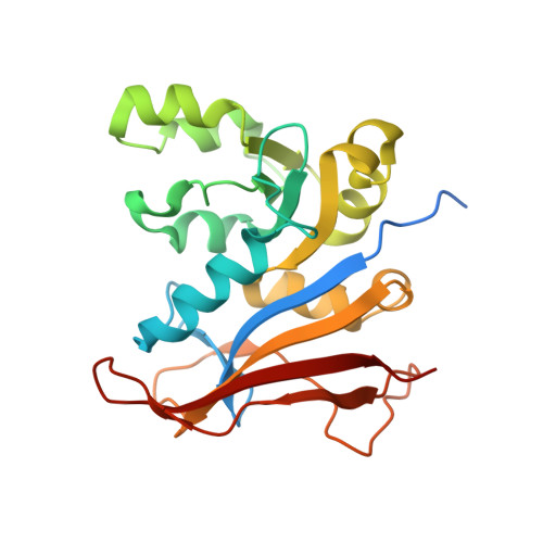
    Dihydrofolate reductase (DHFR) is a potential drug target for Trypanosoma brucei, a human parasite, which is the causative agent for African sleeping sickness. No drug is available against this target, since none of the classical antifolates such as pyrimethamine (PYR), cycloguanil, or trimethoprim are effective as selective inhibitors of T. brucei DHFR (TbDHFR). In order to design effective drugs that target TbDHFR, co-crystal structures with bound antifolates were studied. On comparison with malarial Plasmodium falciparum DHFR (PfDHFR), the co-crystal structures of wild-type TbDHFR reveal greater structural similarities to a mutant PfDHFR causing antifolate resistance than the wild-type enzyme. TbDHFR imposes steric hindrance for rigid inhibitors like PYR around Thr86, which is equivalent to Ser108Asn of the malarial enzymes. In addition, a missing residue on TbDHFR active-site loop together with the presence of Ile51 widens its active site even further than the structural effect of Asn51Ile, which is observed in PfDHFR structures. The structural similarities are paralleled by the similarly poor affinities of the trypanosomal enzyme for rigid inhibitors. Mutations of TbDHFR at Thr86 resulted in 10-fold enhancement or 7-fold reduction in the rigid inhibitors affinities for Thr86Ser or Thr86Asn, respectively. The co-crystal structure of TbDHFR with a flexible antifolate WR99210 suggests that its greater affinity result from its ability to avoid such Thr86 clash and occupy the widened binding space similarly to what is observed in the PfDHFR structures. Natural resistance to antifolates of TbDHFR can therefore be explained, and potential antifolate chemotherapy of trypanosomiasis should be possible taking this into account.


  • Organizational Affiliation

    National Center for Genetic Engineering and Biotechnology, National Science and Technology Development Agency, Klong Luang, Pathumthani, Thailand.


Macromolecules
Find similar proteins by:  (by identity cutoff)  |  3D Structure
Entity ID: 1
MoleculeChains Sequence LengthOrganismDetailsImage
Bifunctional dihydrofolate reductase-thymidylate synthase
A, B
240Trypanosoma brucei rhodesienseMutation(s): 0 
Gene Names: DHFR
EC: 1.5.1.3
UniProt
Find proteins for Q27783 (Trypanosoma brucei brucei)
Explore Q27783 
Go to UniProtKB:  Q27783
Entity Groups  
Sequence Clusters30% Identity50% Identity70% Identity90% Identity95% Identity100% Identity
UniProt GroupQ27783
Sequence Annotations
Expand
  • Reference Sequence
Small Molecules
Ligands 2 Unique
IDChains Name / Formula / InChI Key2D Diagram3D Interactions
NDP
Query on NDP

Download Ideal Coordinates CCD File 
C [auth A],
E [auth B]
NADPH DIHYDRO-NICOTINAMIDE-ADENINE-DINUCLEOTIDE PHOSPHATE
C21 H30 N7 O17 P3
ACFIXJIJDZMPPO-NNYOXOHSSA-N
WRA
Query on WRA

Download Ideal Coordinates CCD File 
D [auth A],
F [auth B]
6,6-DIMETHYL-1-[3-(2,4,5-TRICHLOROPHENOXY)PROPOXY]-1,6-DIHYDRO-1,3,5-TRIAZINE-2,4-DIAMINE
C14 H18 Cl3 N5 O2
MJZJYWCQPMNPRM-UHFFFAOYSA-N
Binding Affinity Annotations 
IDSourceBinding Affinity
WRA Binding MOAD:  3RG9 Ki: 1.1 (nM) from 1 assay(s)
Experimental Data & Validation

Experimental Data

  • Method: X-RAY DIFFRACTION
  • Resolution: 2.00 Å
  • R-Value Free: 0.221 
  • R-Value Work: 0.183 
  • R-Value Observed: 0.183 
  • Space Group: P 41
Unit Cell:
Length ( Å )Angle ( ˚ )
a = 58.409α = 90
b = 58.409β = 90
c = 151.123γ = 90
Software Package:
Software NamePurpose
HKL-2000data collection
AMoREphasing
CNSrefinement
HKL-2000data reduction
HKL-2000data scaling

Structure Validation

View Full Validation Report


Ligand Structure Quality Assessment 


Entry History 

Deposition Data

Revision History  (Full details and data files)

  • Version 1.0: 2011-06-29
    Type: Initial release
  • Version 1.1: 2011-07-13
    Changes: Version format compliance
  • Version 1.2: 2013-06-26
    Changes: Database references, Other