Pb2+ as modulator of protein-membrane interactions.
Morales, K.A., Lasagna, M., Gribenko, A.V., Yoon, Y., Reinhart, G.D., Lee, J.C., Cho, W., Li, P., Igumenova, T.I.(2011) J Am Chem Soc 133: 10599-10611
- PubMed: 21615172
- DOI: https://doi.org/10.1021/ja2032772
- Primary Citation of Related Structures:
3RDJ, 3TWY - PubMed Abstract:
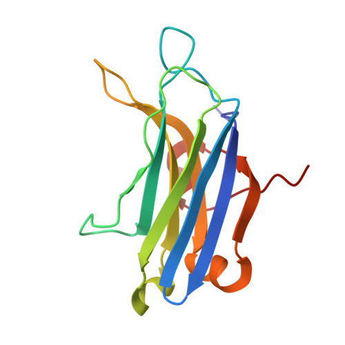
Lead is a potent environmental toxin that mimics the effects of divalent metal ions, such as zinc and calcium, in the context of specific molecular targets and signaling processes. The molecular mechanism of lead toxicity remains poorly understood. The objective of this work was to characterize the effect of Pb(2+) on the structure and membrane-binding properties of C2α. C2α is a peripheral membrane-binding domain of Protein Kinase Cα (PKCα), which is a well-documented molecular target of lead. Using NMR and isothermal titration calorimetry (ITC) techniques, we established that C2α binds Pb(2+) with higher affinity than its natural cofactor, Ca(2+). To gain insight into the coordination geometry of protein-bound Pb(2+), we determined the crystal structures of apo and Pb(2+)-bound C2α at 1.9 and 1.5 Å resolution, respectively. A comparison of these structures revealed that the metal-binding site is not preorganized and that rotation of the oxygen-donating side chains is required for the metal coordination to occur. Remarkably, we found that holodirected and hemidirected coordination geometries for the two Pb(2+) ions coexist within a single protein molecule. Using protein-to-membrane Förster resonance energy transfer (FRET) spectroscopy, we demonstrated that Pb(2+) displaces Ca(2+) from C2α in the presence of lipid membranes through the high-affinity interaction with the membrane-unbound C2α. In addition, Pb(2+) associates with phosphatidylserine-containing membranes and thereby competes with C2α for the membrane-binding sites. This process can contribute to the inhibitory effect of Pb(2+) on the PKCα activity.
Organizational Affiliation:
Department of Biochemistry and Biophysics, Texas A&M University, College Station, Texas 77843, USA.