3RAS

Crystal structure of 1-deoxy-D-xylulose 5-phosphate reductoisomerase (DXR) complexed with a lipophilic phosphonate inhibitor


Experimental Data Snapshot

  • Method: X-RAY DIFFRACTION
  • Resolution: 2.55 Å
  • R-Value Free: 0.278 
  • R-Value Work: 0.208 
  • R-Value Observed: 0.208 

wwPDB Validation   3D Report Full Report


Ligand Structure Quality Assessment 


This is version 1.3 of the entry. See complete history


Literature

Inhibition of 1-deoxy-D-xylulose-5-phosphate reductoisomerase by lipophilic phosphonates: SAR, QSAR, and crystallographic studies.

Deng, L.Diao, J.Chen, P.Pujari, V.Yao, Y.Cheng, G.Crick, D.C.Prasad, B.V.Song, Y.

(2011) J Med Chem 54: 4721-4734

  • DOI: https://doi.org/10.1021/jm200363d
  • Primary Citation of Related Structures:  
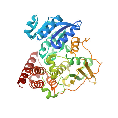
    3RAS

  • PubMed Abstract: 

    1-Deoxy-D-xylulose-5-phosphate reductoisomerase (DXR) is a novel target for developing new antibacterial (including antituberculosis) and antimalaria drugs. Forty-one lipophilic phosphonates, representing a new class of DXR inhibitors, were synthesized, among which 5-phenylpyridin-2-ylmethylphosphonic acid possesses the most activity against E. coli DXR (EcDXR) with a K(i) of 420 nM. Structure-activity relationships (SAR) are discussed, which can be rationalized using our EcDXR:inhibitor structures, and a predictive quantitative SAR (QSAR) model is also developed. Since inhibition studies of DXR from Mycobacterium tuberculosis (MtDXR) have not been performed well, 48 EcDXR inhibitors with a broad chemical diversity were found, however, to generally exhibit considerably reduced activity against MtDXR. The crystal structure of a MtDXR:inhibitor complex reveals the flexible loop containing the residues 198-208 has no strong interactions with the 3,4-dichlorophenyl group of the inhibitor, representing a structural basis for the reduced activity. Overall, these results provide implications in the future design and development of potent DXR inhibitors.


  • Organizational Affiliation

    Department of Pharmacology, Baylor College of Medicine, Houston, Texas 77030, United States.


Macromolecules
Find similar proteins by:  (by identity cutoff)  |  3D Structure
Entity ID: 1
MoleculeChains Sequence LengthOrganismDetailsImage
1-deoxy-D-xylulose 5-phosphate reductoisomerase
A, B
398Mycobacterium tuberculosisMutation(s): 0 
Gene Names: dxrRv2870cMT2938MTCY274.01c
EC: 1.1.1.267
UniProt
Find proteins for P9WNS1 (Mycobacterium tuberculosis (strain ATCC 25618 / H37Rv))
Explore P9WNS1 
Go to UniProtKB:  P9WNS1
Entity Groups  
Sequence Clusters30% Identity50% Identity70% Identity90% Identity95% Identity100% Identity
UniProt GroupP9WNS1
Sequence Annotations
Expand
  • Reference Sequence
Small Molecules
Ligands 5 Unique
IDChains Name / Formula / InChI Key2D Diagram3D Interactions
NDP
Query on NDP

Download Ideal Coordinates CCD File 
C [auth A],
G [auth B]
NADPH DIHYDRO-NICOTINAMIDE-ADENINE-DINUCLEOTIDE PHOSPHATE
C21 H30 N7 O17 P3
ACFIXJIJDZMPPO-NNYOXOHSSA-N
DCV
Query on DCV

Download Ideal Coordinates CCD File 
E [auth A],
I [auth B]
[(1R)-3-[acetyl(hydroxy)amino]-1-(3,4-dichlorophenyl)propyl]phosphonic acid
C11 H14 Cl2 N O5 P
ABGCTQYLJZGMBM-LLVKDONJSA-N
FM5
Query on FM5

Download Ideal Coordinates CCD File 
D [auth A],
H [auth B]
3-(N-HYDROXYACETAMIDO)-1-(3,4-DICHLOROPHENYL)PROPYLPHOSPHONIC ACID
C11 H14 Cl2 N O5 P
ABGCTQYLJZGMBM-NSHDSACASA-N
SO4
Query on SO4

Download Ideal Coordinates CCD File 
K [auth B]SULFATE ION
O4 S
QAOWNCQODCNURD-UHFFFAOYSA-L
MN
Query on MN

Download Ideal Coordinates CCD File 
F [auth A],
J [auth B]
MANGANESE (II) ION
Mn
WAEMQWOKJMHJLA-UHFFFAOYSA-N
Binding Affinity Annotations 
IDSourceBinding Affinity
DCV BindingDB:  3RAS Ki: 1500 (nM) from 1 assay(s)
Kd: 360 (nM) from 1 assay(s)
Experimental Data & Validation

Experimental Data

  • Method: X-RAY DIFFRACTION
  • Resolution: 2.55 Å
  • R-Value Free: 0.278 
  • R-Value Work: 0.208 
  • R-Value Observed: 0.208 
  • Space Group: P 1 21 1
Unit Cell:
Length ( Å )Angle ( ˚ )
a = 67.25α = 90
b = 65.179β = 101.28
c = 85.408γ = 90
Software Package:
Software NamePurpose
StructureStudiodata collection
PHASERphasing
CNSrefinement
HKL-2000data reduction
HKL-2000data scaling

Structure Validation

View Full Validation Report


Ligand Structure Quality Assessment 


Entry History 

Deposition Data

Revision History  (Full details and data files)

  • Version 1.0: 2011-05-25
    Type: Initial release
  • Version 1.1: 2011-07-13
    Changes: Version format compliance
  • Version 1.2: 2012-07-25
    Changes: Database references
  • Version 1.3: 2024-02-21
    Changes: Data collection, Database references, Derived calculations