3RAB

GPPNHP-BOUND RAB3A AT 2.0 A RESOLUTION


Experimental Data Snapshot

  • Method: X-RAY DIFFRACTION
  • Resolution: 2.00 Å
  • R-Value Free: 0.237 
  • R-Value Work: 0.191 

wwPDB Validation   3D Report Full Report


Ligand Structure Quality Assessment 


This is version 1.3 of the entry. See complete history


Literature

Structural basis of activation and GTP hydrolysis in Rab proteins.

Dumas, J.J.Zhu, Z.Connolly, J.L.Lambright, D.G.

(1999) Structure 7: 413-423

  • DOI: https://doi.org/10.1016/s0969-2126(99)80054-9
  • Primary Citation of Related Structures:  
    3RAB

  • PubMed Abstract: 

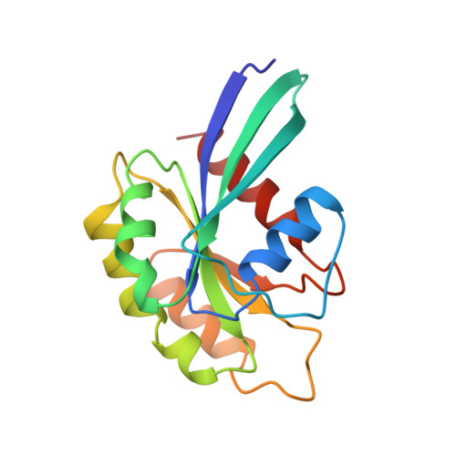
    Rab proteins comprise a large family of GTPases that regulate vesicle trafficking. Despite conservation of critical residues involved in nucleotide binding and hydrolysis, Rab proteins exhibit low sequence identity with other GTPases, and the structural basis for Rab function remains poorly characterized.


  • Organizational Affiliation

    Program in Molecular Medicine, University of Massachusetts Medical Center, 373 Plantation Street, Worcester, MA 01605, USA.


Macromolecules
Find similar proteins by:  (by identity cutoff)  |  3D Structure
Entity ID: 1
MoleculeChains Sequence LengthOrganismDetailsImage
PROTEIN (RAB3A)169Rattus norvegicusMutation(s): 0 
UniProt
Find proteins for P63012 (Rattus norvegicus)
Explore P63012 
Go to UniProtKB:  P63012
Entity Groups  
Sequence Clusters30% Identity50% Identity70% Identity90% Identity95% Identity100% Identity
UniProt GroupP63012
Sequence Annotations
Expand
  • Reference Sequence
Small Molecules
Ligands 2 Unique
IDChains Name / Formula / InChI Key2D Diagram3D Interactions
GNP
Query on GNP

Download Ideal Coordinates CCD File 
C [auth A]PHOSPHOAMINOPHOSPHONIC ACID-GUANYLATE ESTER
C10 H17 N6 O13 P3
UQABYHGXWYXDTK-UUOKFMHZSA-N
MG
Query on MG

Download Ideal Coordinates CCD File 
B [auth A]MAGNESIUM ION
Mg
JLVVSXFLKOJNIY-UHFFFAOYSA-N
Experimental Data & Validation

Experimental Data

  • Method: X-RAY DIFFRACTION
  • Resolution: 2.00 Å
  • R-Value Free: 0.237 
  • R-Value Work: 0.191 
  • Space Group: C 1 2 1
Unit Cell:
Length ( Å )Angle ( ˚ )
a = 88.9α = 90
b = 35β = 107.2
c = 58.6γ = 90
Software Package:
Software NamePurpose
AMoREphasing
X-PLORrefinement
DENZOdata reduction
SCALEPACKdata scaling

Structure Validation

View Full Validation Report


Ligand Structure Quality Assessment 


Entry History 

Deposition Data

Revision History  (Full details and data files)

  • Version 1.0: 1999-04-16
    Type: Initial release
  • Version 1.1: 2008-04-26
    Changes: Version format compliance
  • Version 1.2: 2011-07-13
    Changes: Version format compliance
  • Version 1.3: 2023-09-13
    Changes: Data collection, Database references, Derived calculations, Refinement description