Biochemical and structural characterization of Plasmodium falciparum glutamate dehydrogenase 2.
Zocher, K., Fritz-Wolf, K., Kehr, S., Fischer, M., Rahlfs, S., Becker, K.(2012) Mol Biochem Parasitol 183: 52-62
- PubMed: 22342964
- DOI: https://doi.org/10.1016/j.molbiopara.2012.01.007
- Primary Citation of Related Structures:
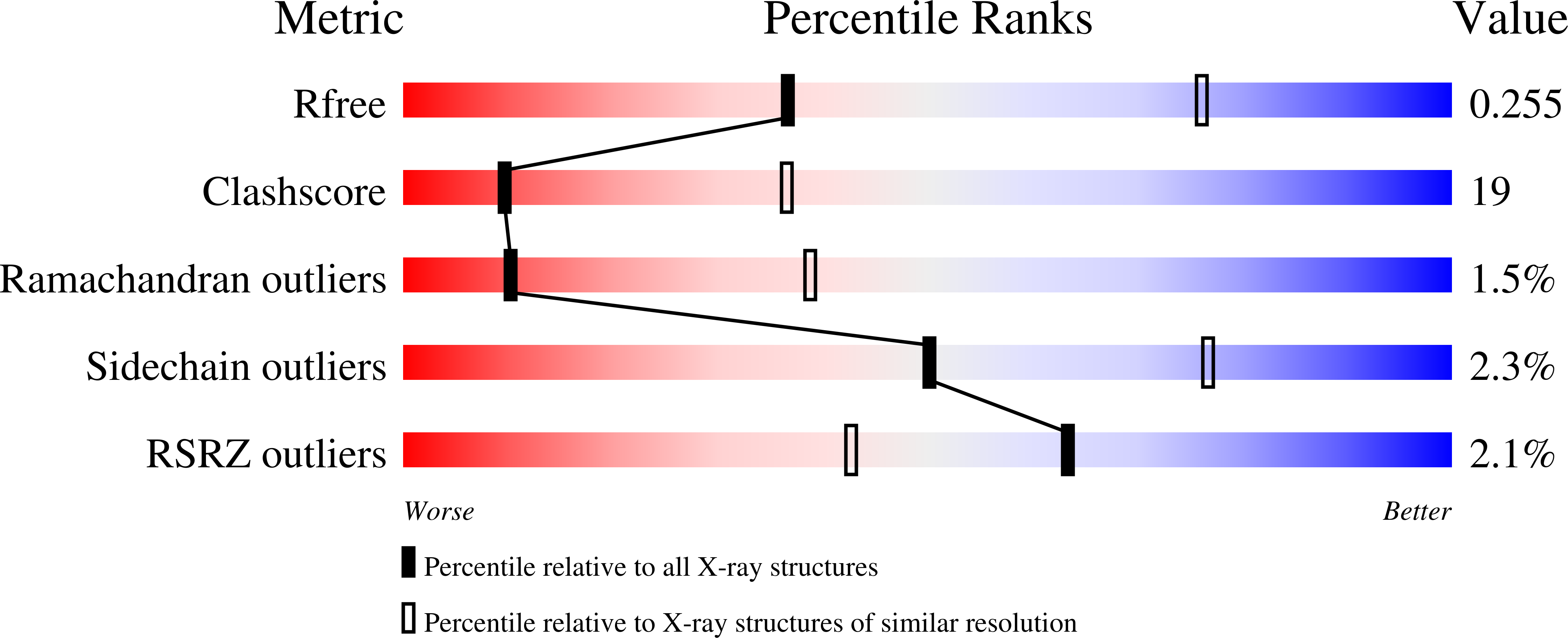
3R3J - PubMed Abstract:
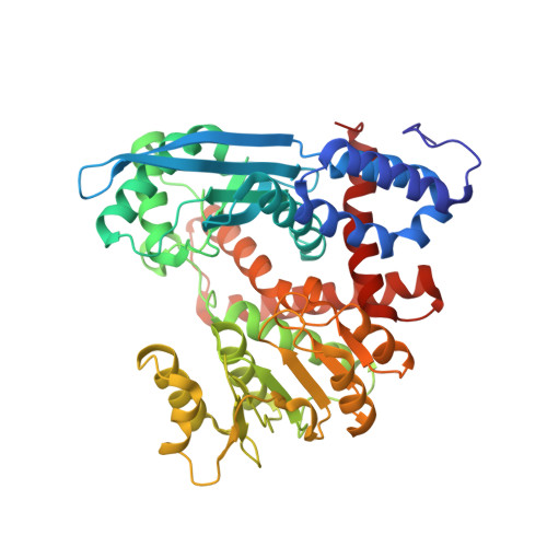
Glutamate dehydrogenases (GDHs) play key roles in cellular redox, amino acid, and energy metabolism, thus representing potential targets for pharmacological interventions. Here we studied the functional network provided by the three known glutamate dehydrogenases of the malaria parasite Plasmodium falciparum. The recombinant production of the previously described PfGDH1 as hexahistidyl-tagged proteins was optimized. Additionally, PfGDH2 was cloned, recombinantly produced, and characterized. Like PfGDH1, PfGDH2 is an NADP(H)-dependent enzyme with a specific activity comparable to PfGDH1 but with slightly higher K(m) values for its substrates. The three-dimensional structure of hexameric PfGDH2 was solved to 3.1 Å resolution. The overall structure shows high similarity with PfGDH1 but with significant differences occurring at the subunit interface. As in mammalian GDH1, in PfGDH2 the subunit-subunit interactions are mainly assisted by hydrogen bonds and hydrophobic interactions, whereas in PfGDH1 these contacts are mediated by networks of salt bridges and hydrogen bonds. In accordance with this, the known bovine GDH inhibitors hexachlorophene, GW5074, and bithionol were more effective on PfGDH2 than on PfGDH1. Subcellular localization was determined for all three plasmodial GDHs by fusion with the green fluorescent protein. Based on our data, PfGDH1 and PfGDH3 are cytosolic proteins whereas PfGDH2 clearly localizes to the apicoplast, a plastid-like organelle specific for apicomplexan parasites. This study provides new insights into the structure and function of GDH isoenzymes of P. falciparum, which represent potential targets for the development of novel antimalarial drugs.
Organizational Affiliation:
Interdisciplinary Research Center, Justus Liebig University, Heinrich-Buff-Ring 26-32, D-35392 Giessen, Germany. kathleen.zocher@gmx.de