3R0H

Structure of INAD PDZ45 in complex with NG2 peptide


Experimental Data Snapshot

  • Method: X-RAY DIFFRACTION
  • Resolution: 2.60 Å
  • R-Value Free: 0.263 
  • R-Value Work: 0.198 
  • R-Value Observed: 0.202 

wwPDB Validation   3D Report Full Report


Ligand Structure Quality Assessment 


This is version 1.1 of the entry. See complete history


Literature

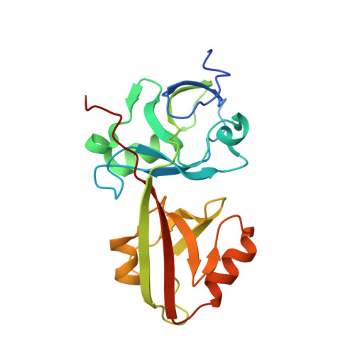
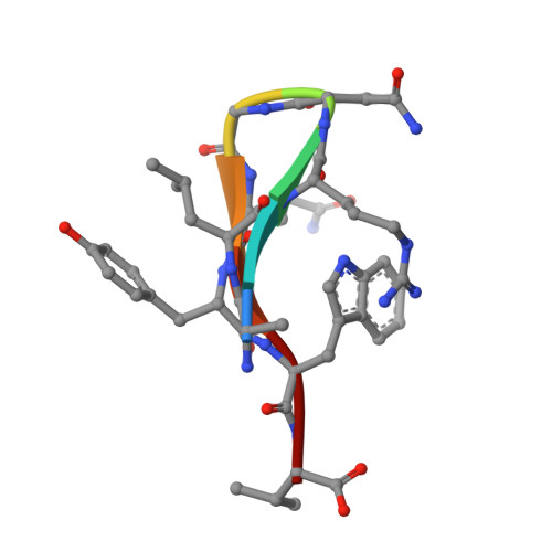
The INAD scaffold is a dynamic, redox-regulated modulator of signaling in the Drosophila eye

Liu, W.Wen, W.Wei, Z.Yu, J.Ye, F.Liu, C.-H.Hardie, R.C.Zhang, M.

(2011) Cell 145: 1088-1101

  • DOI: https://doi.org/10.1016/j.cell.2011.05.015
  • Primary Citation of Related Structures:  
    2LA8, 3R0H

  • PubMed Abstract: 

    INAD is a scaffolding protein that regulates signaling in Drosophila photoreceptors. One of its PDZ domains, PDZ5, cycles between reduced and oxidized forms in response to light, but it is unclear how light affects its redox potential. Through biochemical and structural studies, we show that the redox potential of PDZ5 is allosterically regulated by its interaction with another INAD domain, PDZ4. Whereas isolated PDZ5 is stable in the oxidized state, formation of a PDZ45 "supramodule" locks PDZ5 in the reduced state by raising the redox potential of its Cys606/Cys645 disulfide bond by ∼330 mV. Acidification, potentially mediated via light and PLCβ-mediated hydrolysis of PIP(2), disrupts the interaction between PDZ4 and PDZ5, leading to PDZ5 oxidation and dissociation from the TRP Ca(2+) channel, a key component of fly visual signaling. These results show that scaffolding proteins can actively modulate the intrinsic redox potentials of their disulfide bonds to exert regulatory roles in signaling.


  • Organizational Affiliation

    Division of Life Science, Molecular Neuroscience Center, State Key Laboratory of Molecular Neuroscience, Hong Kong University of Science and Technology, Clear Water Bay, Kowloon, Hong Kong.


Macromolecules
Find similar proteins by:  (by identity cutoff)  |  3D Structure
Entity ID: 1
MoleculeChains Sequence LengthOrganismDetailsImage
Inactivation-no-after-potential D protein206Drosophila melanogasterMutation(s): 0 
Gene Names: CG3504inaD
UniProt
Find proteins for Q24008 (Drosophila melanogaster)
Explore Q24008 
Go to UniProtKB:  Q24008
Entity Groups  
Sequence Clusters30% Identity50% Identity70% Identity90% Identity95% Identity100% Identity
UniProt GroupQ24008
Sequence Annotations
Expand
  • Reference Sequence

Find similar proteins by:  Sequence   |   3D Structure  

Entity ID: 2
MoleculeChains Sequence LengthOrganismDetailsImage
NG29N/AMutation(s): 0 
Sequence Annotations
Expand
  • Reference Sequence
Small Molecules
Ligands 3 Unique
IDChains Name / Formula / InChI Key2D Diagram3D Interactions
DTV
Query on DTV

Download Ideal Coordinates CCD File 
R [auth B](2S,3S)-1,4-DIMERCAPTOBUTANE-2,3-DIOL
C4 H10 O2 S2
VHJLVAABSRFDPM-QWWZWVQMSA-N
DTT
Query on DTT

Download Ideal Coordinates CCD File 
Q [auth A],
S [auth B],
U [auth D],
V [auth D]
2,3-DIHYDROXY-1,4-DITHIOBUTANE
C4 H10 O2 S2
VHJLVAABSRFDPM-IMJSIDKUSA-N
SO4
Query on SO4

Download Ideal Coordinates CCD File 
T [auth C],
W [auth G],
X [auth G],
Y [auth H],
Z [auth H]
SULFATE ION
O4 S
QAOWNCQODCNURD-UHFFFAOYSA-L
Experimental Data & Validation

Experimental Data

  • Method: X-RAY DIFFRACTION
  • Resolution: 2.60 Å
  • R-Value Free: 0.263 
  • R-Value Work: 0.198 
  • R-Value Observed: 0.202 
  • Space Group: P 21 21 21
Unit Cell:
Length ( Å )Angle ( ˚ )
a = 73.079α = 90
b = 134.988β = 90
c = 215.986γ = 90
Software Package:
Software NamePurpose
SCALEPACKdata scaling
REFMACrefinement
PDB_EXTRACTdata extraction
HKL-2000data reduction
HKL-2000data scaling
SHARPphasing

Structure Validation

View Full Validation Report


Ligand Structure Quality Assessment 


Entry History 

Deposition Data

Revision History  (Full details and data files)

  • Version 1.0: 2011-11-30
    Type: Initial release
  • Version 1.1: 2024-03-20
    Changes: Data collection, Database references, Derived calculations