


Crystal structure of Cytosine Deaminase from Escherichia Coli complexed with two zinc atoms in the active site
Fedorov, A.A., Fedorov, E.V., Kamat, S., Hitchcock, D., Raushel, F.M., Almo, S.C.To be published.
Experimental Data Snapshot
Entity ID: 1 | |||||
|---|---|---|---|---|---|
| Molecule | Chains | Sequence Length | Organism | Details | Image |
| Cytosine deaminase | 427 | Escherichia coli K-12 | Mutation(s): 0 Gene Names: codA, b0337, JW0328 EC: 3.5.4.1 | 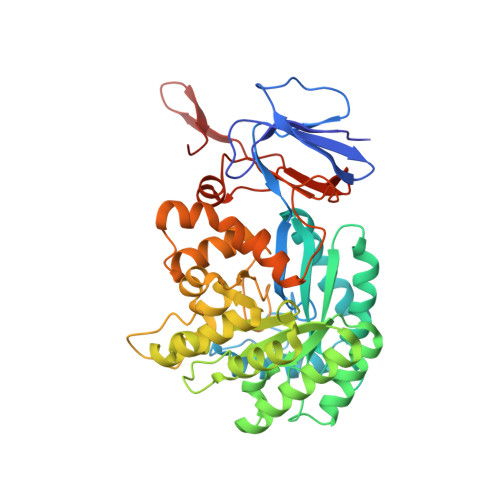 | |
UniProt | |||||
Find proteins for P25524 (Escherichia coli (strain K12)) Explore P25524 Go to UniProtKB: P25524 | |||||
Entity Groups | |||||
| Sequence Clusters | 30% Identity50% Identity70% Identity90% Identity95% Identity100% Identity | ||||
| UniProt Group | P25524 | ||||
Sequence AnnotationsExpand | |||||
| |||||
| Ligands 3 Unique | |||||
|---|---|---|---|---|---|
| ID | Chains | Name / Formula / InChI Key | 2D Diagram | 3D Interactions | |
| PXN Query on PXN | B [auth A] | (2S)-1-[3-{[(2R)-2-hydroxypropyl]oxy}-2,2-bis({[(2R)-2-hydroxypropyl]oxy}methyl)propoxy]propan-2-ol C17 H36 O8 GXEZGLLPFFKHGE-FPCVCCKLSA-N |  | ||
| GOL Query on GOL | F [auth A], G [auth A] | GLYCEROL C3 H8 O3 PEDCQBHIVMGVHV-UHFFFAOYSA-N |  | ||
| ZN Query on ZN | C [auth A], D [auth A], E [auth A] | ZINC ION Zn PTFCDOFLOPIGGS-UHFFFAOYSA-N |  | ||
| Length ( Å ) | Angle ( ˚ ) |
|---|---|
| a = 145.492 | α = 90 |
| b = 145.492 | β = 90 |
| c = 199.704 | γ = 120 |
| Software Name | Purpose |
|---|---|
| ADSC | data collection |
| BALBES | phasing |
| PHENIX | refinement |
| DENZO | data reduction |
| SCALEPACK | data scaling |
RCSB PDB (citation) is hosted by
RCSB PDB is a member of the

