3QYO

Sensitivity of receptor internal motions to ligand binding affinity and kinetic off-rate


Experimental Data Snapshot

  • Method: X-RAY DIFFRACTION
  • Resolution: 2.09 Å
  • R-Value Free: 0.236 
  • R-Value Work: 0.165 
  • R-Value Observed: 0.169 

wwPDB Validation   3D Report Full Report


Ligand Structure Quality Assessment 


This is version 1.3 of the entry. See complete history


Literature

Evidence for dynamics in proteins as a mechanism for ligand dissociation.

Carroll, M.J.Mauldin, R.V.Gromova, A.V.Singleton, S.F.Collins, E.J.Lee, A.L.

(2012) Nat Chem Biol 8: 246-252

  • DOI: https://doi.org/10.1038/nchembio.769
  • Primary Citation of Related Structures:  
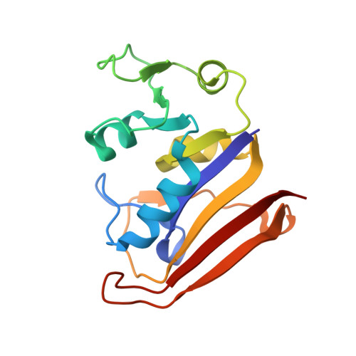
    3QYL, 3QYO, 3R33

  • PubMed Abstract: 

    Signal transduction, regulatory processes and pharmaceutical responses are highly dependent upon ligand residence times. Gaining insight into how physical factors influence residence times (1/k(off)) should enhance our ability to manipulate biological interactions. We report experiments that yield structural insight into k(off) involving a series of eight 2,4-diaminopyrimidine inhibitors of dihydrofolate reductase whose binding affinities vary by six orders of magnitude. NMR relaxation-dispersion experiments revealed a common set of residues near the binding site that undergo a concerted millisecond-timescale switching event to a previously unidentified conformation. The rate of switching from ground to excited conformations correlates exponentially with the binding affinity K(i) and k(off), suggesting that protein dynamics serves as a mechanical initiator of ligand dissociation within this series and potentially for other macromolecule-ligand systems. Although the forward rate of conformational exchange, k(conf,forward), is faster than k(off), the use of the ligand series allowed for connections to be drawn between kinetic events on different timescales.


  • Organizational Affiliation

    Division of Chemical Biology and Medicinal Chemistry, Eshelman School of Pharmacy, University of North Carolina at Chapel Hill, Chapel Hill, North Carolina, USA.


Macromolecules
Find similar proteins by:  (by identity cutoff)  |  3D Structure
Entity ID: 1
MoleculeChains Sequence LengthOrganismDetailsImage
Dihydrofolate reductase159Escherichia coli K-12Mutation(s): 0 
EC: 1.5.1.3
UniProt
Find proteins for P0ABQ4 (Escherichia coli (strain K12))
Explore P0ABQ4 
Go to UniProtKB:  P0ABQ4
Entity Groups  
Sequence Clusters30% Identity50% Identity70% Identity90% Identity95% Identity100% Identity
UniProt GroupP0ABQ4
Sequence Annotations
Expand
  • Reference Sequence
Small Molecules
Ligands 3 Unique
IDChains Name / Formula / InChI Key2D Diagram3D Interactions
NDP
Query on NDP

Download Ideal Coordinates CCD File 
C [auth A]NADPH DIHYDRO-NICOTINAMIDE-ADENINE-DINUCLEOTIDE PHOSPHATE
C21 H30 N7 O17 P3
ACFIXJIJDZMPPO-NNYOXOHSSA-N
Q24
Query on Q24

Download Ideal Coordinates CCD File 
B [auth A]quinazoline-2,4-diamine
C8 H8 N4
XELRMPRLCPFTBH-UHFFFAOYSA-N
CA
Query on CA

Download Ideal Coordinates CCD File 
D [auth A]
E [auth A]
F [auth A]
G [auth A]
H [auth A]
D [auth A],
E [auth A],
F [auth A],
G [auth A],
H [auth A],
I [auth A],
J [auth A],
K [auth A]
CALCIUM ION
Ca
BHPQYMZQTOCNFJ-UHFFFAOYSA-N
Binding Affinity Annotations 
IDSourceBinding Affinity
Q24 Binding MOAD:  3QYO Ki: 7000 (nM) from 1 assay(s)
Experimental Data & Validation

Experimental Data

  • Method: X-RAY DIFFRACTION
  • Resolution: 2.09 Å
  • R-Value Free: 0.236 
  • R-Value Work: 0.165 
  • R-Value Observed: 0.169 
  • Space Group: P 21 21 21
Unit Cell:
Length ( Å )Angle ( ˚ )
a = 33.99α = 90
b = 45.11β = 90
c = 97.8γ = 90
Software Package:
Software NamePurpose
HKL-2000data collection
PHASERphasing
BUSTERrefinement
MOSFLMdata reduction
SCALAdata scaling

Structure Validation

View Full Validation Report


Ligand Structure Quality Assessment 


Entry History 

Deposition Data

Revision History  (Full details and data files)

  • Version 1.0: 2012-01-18
    Type: Initial release
  • Version 1.1: 2012-04-25
    Changes: Database references
  • Version 1.2: 2014-11-12
    Changes: Structure summary
  • Version 1.3: 2024-02-21
    Changes: Data collection, Database references, Derived calculations