3QW9

Crystal structure of betaglycan ZP-C domain


Experimental Data Snapshot

  • Method: X-RAY DIFFRACTION
  • Resolution: 2.00 Å
  • R-Value Free: 0.245 
  • R-Value Work: 0.186 
  • R-Value Observed: 0.189 

wwPDB Validation   3D Report Full Report


This is version 2.0 of the entry. See complete history


Literature

Structure of betaglycan zona pellucida (ZP)-C domain provides insights into ZP-mediated protein polymerization and TGF-{beta} binding.

Lin, S.J.Hu, Y.Zhu, J.Woodruff, T.K.Jardetzky, T.S.

(2011) Proc Natl Acad Sci U S A 108: 5232-5236

  • DOI: https://doi.org/10.1073/pnas.1010689108
  • Primary Citation of Related Structures:  
    3QW9

  • PubMed Abstract: 

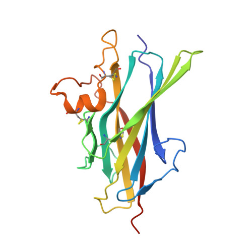
    The zona pellucida (ZP) domain is a bipartite protein structural element comprised of ZP-N and ZP-C regions. Most notable for its ability to mediate protein polymerization, many ZP proteins polymerize and assemble into long fibrils that form specialized extracellular matrices. Other ZP proteins (namely, betaglycan and endoglin) do not polymerize but serve as important membrane coreceptors for ligands in the transforming growth factor-β (TGF-β) superfamily. Here, we present the 2.0-Å resolution crystal structure of the betaglycan ZP-C region in combination with a downstream region known as the external hydrophobic patch (EHP). Similar to the ZP-N region, the ZP-C region also adopts an immunoglobulin-like fold, despite sharing no sequence homology and possessing different disulfide linkages. The EHP region, which was previously thought to be external to the ZP region, is integral to the ZP-C domain and corresponds to the ZP-C G strand. Our structure also indicates that the critical maturation cleavage of ZP proteins, a process that activates nascent ZP proteins for polymerization, occurs within the immunoglobulin domain at the FG loop. Nonpolymerizing ZP proteins such as betaglycan and endoglin do not contain this cleavage site. Finally, our structure suggests that the AB loop and the convex surface pocket are regions important for TGF-β ligand binding.


  • Organizational Affiliation

    Department of Structural Biology, Stanford University School of Medicine, 371 Serra Mall, Stanford, CA 94305, USA.


Macromolecules
Find similar proteins by:  (by identity cutoff)  |  3D Structure
Entity ID: 1
MoleculeChains Sequence LengthOrganismDetailsImage
Transforming growth factor beta receptor type 3
A, B
176Rattus norvegicusMutation(s): 0 
Gene Names: Tgfbr3
UniProt
Find proteins for P26342 (Rattus norvegicus)
Explore P26342 
Go to UniProtKB:  P26342
Entity Groups  
Sequence Clusters30% Identity50% Identity70% Identity90% Identity95% Identity100% Identity
UniProt GroupP26342
Sequence Annotations
Expand
  • Reference Sequence
Oligosaccharides

Help

Entity ID: 2
MoleculeChains Length2D Diagram Glycosylation3D Interactions
beta-D-mannopyranose-(1-4)-2-acetamido-2-deoxy-beta-D-glucopyranose-(1-4)-[alpha-L-fucopyranose-(1-3)][alpha-L-fucopyranose-(1-6)]2-acetamido-2-deoxy-beta-D-glucopyranose
C
5N-Glycosylation
Glycosylation Resources
GlyTouCan:  G92953ND
GlyCosmos:  G92953ND
Entity ID: 3
MoleculeChains Length2D Diagram Glycosylation3D Interactions
alpha-D-mannopyranose-(1-3)-[alpha-D-mannopyranose-(1-6)]beta-D-mannopyranose-(1-4)-2-acetamido-2-deoxy-beta-D-glucopyranose-(1-4)-[alpha-L-fucopyranose-(1-3)][alpha-L-fucopyranose-(1-6)]2-acetamido-2-deoxy-beta-D-glucopyranose
D
7N-Glycosylation
Glycosylation Resources
GlyTouCan:  G11460AB
GlyCosmos:  G11460AB
GlyGen:  G11460AB
Experimental Data & Validation

Experimental Data

  • Method: X-RAY DIFFRACTION
  • Resolution: 2.00 Å
  • R-Value Free: 0.245 
  • R-Value Work: 0.186 
  • R-Value Observed: 0.189 
  • Space Group: P 21 21 21
Unit Cell:
Length ( Å )Angle ( ˚ )
a = 53.527α = 90
b = 63.574β = 90
c = 107.217γ = 90
Software Package:
Software NamePurpose
DENZOdata reduction
SCALEPACKdata scaling
SHARPphasing
REFMACrefinement
PDB_EXTRACTdata extraction

Structure Validation

View Full Validation Report


Entry History 

Deposition Data

Revision History  (Full details and data files)

  • Version 1.0: 2011-04-06
    Type: Initial release
  • Version 1.1: 2011-07-13
    Changes: Version format compliance
  • Version 2.0: 2020-07-29
    Type: Remediation
    Reason: Carbohydrate remediation
    Changes: Advisory, Atomic model, Data collection, Database references, Derived calculations, Structure summary