3QT4

Structure of digestive procathepsin L 3 of Tenebrio molitor larval midgut


Experimental Data Snapshot

  • Method: X-RAY DIFFRACTION
  • Resolution: 2.11 Å
  • R-Value Free: 0.201 
  • R-Value Work: 0.158 
  • R-Value Observed: 0.160 

wwPDB Validation   3D Report Full Report


This is version 1.4 of the entry. See complete history


Literature

The 3D structure and function of digestive cathepsin L-like proteinases of Tenebrio molitor larval midgut.

Beton, D.Guzzo, C.R.Ribeiro, A.F.Farah, C.S.Terra, W.R.

(2012) Insect Biochem Mol Biol 42: 655-664

  • DOI: https://doi.org/10.1016/j.ibmb.2012.04.010
  • Primary Citation of Related Structures:  
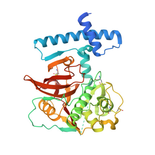
    3QJ3, 3QT4

  • PubMed Abstract: 

    Cathepsin L-like proteinases (CAL) are major digestive proteinases in the beetle Tenebrio molitor. Procathepsin Ls 2 (pCAL2) and 3 (pCAL3) were expressed as recombinant proteins in Escherichia coli, purified and activated under acidic conditions. Immunoblot analyses of different T. molitor larval tissues demonstrated that a polyclonal antibody to pCAL3 recognized pCAL3 and cathepsin L 3 (CAL3) only in the anterior two-thirds of midgut tissue and midgut luminal contents of T. molitor larvae. Furthermore, immunocytolocalization data indicated that pCAL3 occurs in secretory vesicles and microvilli in anterior midgut. Therefore CAL3, like cathepsin L 2 (CAL2), is a digestive enzyme secreted by T. molitor anterior midgut. CAL3 hydrolyses Z-FR-MCA and Z-RR-MCA (typical cathepsin substrates), whereas CAL2 hydrolyses only Z-FR-MCA. Active site mutants (pCAL2C25S and pCAL3C26S) were constructed by replacing the catalytic cysteine with serine to prevent autocatalytic processing. Recombinant pCAL2 and pCAL3 mutants (pCAL2C25S and pCAL3C26S) were prepared, crystallized and their 3D structures determined at 1.85 and 2.1 Å, respectively. While the overall structure of these enzymes is similar to other members of the papain superfamily, structural differences in the S2 subsite explain their substrate specificities. The data also supported models for CAL trafficking to lysosomes and to secretory vesicles to be discharged into midgut contents.


  • Organizational Affiliation

    Departamento de Bioquímica, Instituto de Química, Universidade de São Paulo, C. P. 26077, 05513-970 São Paulo, Brazil.


Macromolecules
Find similar proteins by:  (by identity cutoff)  |  3D Structure
Entity ID: 1
MoleculeChains Sequence LengthOrganismDetailsImage
Cathepsin-L-like midgut cysteine proteinase329Tenebrio molitorMutation(s): 1 
Gene Names: pCAL3
EC: 3.4.22.15
UniProt
Find proteins for Q7YXL2 (Tenebrio molitor)
Explore Q7YXL2 
Go to UniProtKB:  Q7YXL2
Entity Groups  
Sequence Clusters30% Identity50% Identity70% Identity90% Identity95% Identity100% Identity
UniProt GroupQ7YXL2
Sequence Annotations
Expand
  • Reference Sequence
Small Molecules
Ligands 3 Unique
IDChains Name / Formula / InChI Key2D Diagram3D Interactions
PG6
Query on PG6

Download Ideal Coordinates CCD File 
S [auth A],
T [auth A]
1-(2-METHOXY-ETHOXY)-2-{2-[2-(2-METHOXY-ETHOXY]-ETHOXY}-ETHANE
C12 H26 O6
DMDPGPKXQDIQQG-UHFFFAOYSA-N
PG4
Query on PG4

Download Ideal Coordinates CCD File 
L [auth A]
M [auth A]
N [auth A]
O [auth A]
P [auth A]
L [auth A],
M [auth A],
N [auth A],
O [auth A],
P [auth A],
Q [auth A],
R [auth A]
TETRAETHYLENE GLYCOL
C8 H18 O5
UWHCKJMYHZGTIT-UHFFFAOYSA-N
PO4
Query on PO4

Download Ideal Coordinates CCD File 
B [auth A]
C [auth A]
D [auth A]
E [auth A]
F [auth A]
B [auth A],
C [auth A],
D [auth A],
E [auth A],
F [auth A],
G [auth A],
H [auth A],
I [auth A],
J [auth A],
K [auth A]
PHOSPHATE ION
O4 P
NBIIXXVUZAFLBC-UHFFFAOYSA-K
Experimental Data & Validation

Experimental Data

  • Method: X-RAY DIFFRACTION
  • Resolution: 2.11 Å
  • R-Value Free: 0.201 
  • R-Value Work: 0.158 
  • R-Value Observed: 0.160 
  • Space Group: C 1 2 1
Unit Cell:
Length ( Å )Angle ( ˚ )
a = 57.634α = 90
b = 89.322β = 92.5
c = 70.076γ = 90
Software Package:
Software NamePurpose
MAR345data collection
PHASERphasing
REFMACrefinement
HKL-2000data reduction
HKL-2000data scaling

Structure Validation

View Full Validation Report


Entry History 

Deposition Data

Revision History  (Full details and data files)

  • Version 1.0: 2012-02-22
    Type: Initial release
  • Version 1.1: 2012-06-20
    Changes: Database references
  • Version 1.2: 2012-08-22
    Changes: Database references
  • Version 1.3: 2017-11-08
    Changes: Refinement description
  • Version 1.4: 2023-09-13
    Changes: Data collection, Database references, Derived calculations, Refinement description