3QR0

Crystal Structure of S. officinalis PLC21


Experimental Data Snapshot

  • Method: X-RAY DIFFRACTION
  • Resolution: 2.00 Å
  • R-Value Free: 0.208 
  • R-Value Work: 0.176 
  • R-Value Observed: 0.178 

wwPDB Validation   3D Report Full Report


This is version 1.3 of the entry. See complete history


Literature

An autoinhibitory helix in the C-terminal region of phospholipase C-beta mediates Galphaq activation.

Lyon, A.M.Tesmer, V.M.Dhamsania, V.D.Thal, D.M.Gutierrez, J.Chowdhury, S.Suddala, K.C.Northup, J.K.Tesmer, J.J.

(2011) Nat Struct Mol Biol 18: 999-1005

  • DOI: https://doi.org/10.1038/nsmb.2095
  • Primary Citation of Related Structures:  
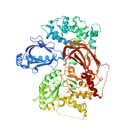
    3QR0, 3QR1

  • PubMed Abstract: 

    The enzyme phospholipase C-β (PLCβ) is a crucial regulator of intracellular calcium levels whose activity is controlled by heptahelical receptors that couple to members of the Gq family of heterotrimeric G proteins. We have determined atomic structures of two invertebrate homologs of PLCβ (PLC21) from cephalopod retina and identified a helix from the C-terminal regulatory region that interacts with a conserved surface of the catalytic core of the enzyme. Mutations designed to disrupt the analogous interaction in human PLCβ3 considerably increase basal activity and diminish stimulation by Gαq. Gαq binding requires displacement of the autoinhibitory helix from the catalytic core, thus providing an allosteric mechanism for activation of PLCβ.


  • Organizational Affiliation

    Life Sciences Institute, Department of Biophysics, University of Michigan, Ann Arbor, Michigan, USA.


Macromolecules
Find similar proteins by:  (by identity cutoff)  |  3D Structure
Entity ID: 1
MoleculeChains Sequence LengthOrganismDetailsImage
phospholipase C-beta (PLC-beta)816Sepia officinalisMutation(s): 0 
UniProt
Find proteins for F2YQ19 (Sepia officinalis)
Explore F2YQ19 
Go to UniProtKB:  F2YQ19
Entity Groups  
Sequence Clusters30% Identity50% Identity70% Identity90% Identity95% Identity100% Identity
UniProt GroupF2YQ19
Sequence Annotations
Expand
  • Reference Sequence
Small Molecules
Ligands 2 Unique
IDChains Name / Formula / InChI Key2D Diagram3D Interactions
GOL
Query on GOL

Download Ideal Coordinates CCD File 
C [auth A]
D [auth A]
E [auth A]
F [auth A]
G [auth A]
C [auth A],
D [auth A],
E [auth A],
F [auth A],
G [auth A],
H [auth A],
I [auth A],
J [auth A],
K [auth A]
GLYCEROL
C3 H8 O3
PEDCQBHIVMGVHV-UHFFFAOYSA-N
CA
Query on CA

Download Ideal Coordinates CCD File 
B [auth A]CALCIUM ION
Ca
BHPQYMZQTOCNFJ-UHFFFAOYSA-N
Experimental Data & Validation

Experimental Data

  • Method: X-RAY DIFFRACTION
  • Resolution: 2.00 Å
  • R-Value Free: 0.208 
  • R-Value Work: 0.176 
  • R-Value Observed: 0.178 
  • Space Group: P 21 21 21
Unit Cell:
Length ( Å )Angle ( ˚ )
a = 60.815α = 90
b = 89.396β = 90
c = 163.051γ = 90
Software Package:
Software NamePurpose
SCALEPACKdata scaling
PHASERphasing
REFMACrefinement
PDB_EXTRACTdata extraction
HKL-2000data collection
DENZOdata reduction

Structure Validation

View Full Validation Report


Entry History 

Deposition Data

Revision History  (Full details and data files)

  • Version 1.0: 2011-08-10
    Type: Initial release
  • Version 1.1: 2011-08-17
    Changes: Database references
  • Version 1.2: 2011-09-14
    Changes: Database references
  • Version 1.3: 2023-09-13
    Changes: Data collection, Database references, Derived calculations, Refinement description