3QPB

Crystal Structure of Streptococcus Pyogenes Uridine Phosphorylase Reveals a Subclass of the NP-I Superfamily


Experimental Data Snapshot

  • Method: X-RAY DIFFRACTION
  • Resolution: 1.82 Å
  • R-Value Free: 0.203 
  • R-Value Work: 0.177 

wwPDB Validation   3D Report Full Report


Ligand Structure Quality Assessment 


This is version 2.1 of the entry. See complete history


Literature

The Crystal Structure of Streptococcus pyogenes Uridine Phosphorylase Reveals a Distinct Subfamily of Nucleoside Phosphorylases.

Tran, T.H.Christoffersen, S.Allan, P.W.Parker, W.B.Serra, I.Terreni, M.Ealick, S.E.

(2011) Biochemistry 50: 6549-6558

  • DOI: https://doi.org/10.1021/bi200707z
  • Primary Citation of Related Structures:  
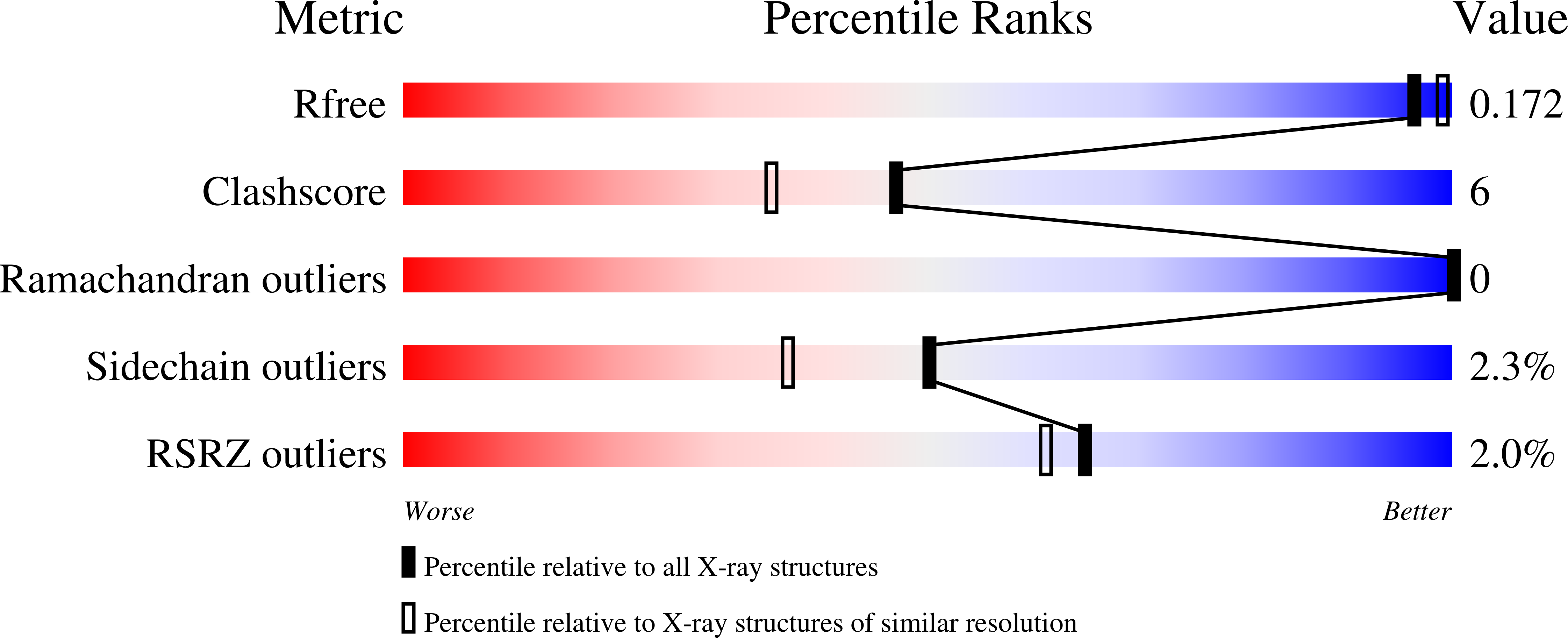
    3QPB

  • PubMed Abstract: 

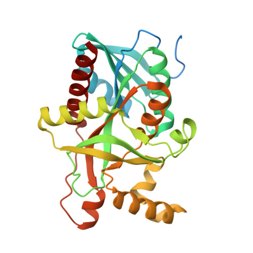
    Uridine phosphorylase (UP), a key enzyme in the pyrimidine salvage pathway, catalyzes the reversible phosphorolysis of uridine or 2'-deoxyuridine to uracil and ribose 1-phosphate or 2'-deoxyribose 1-phosphate. This enzyme belongs to the nucleoside phosphorylase I superfamily whose members show diverse specificity for nucleoside substrates. Phylogenetic analysis shows Streptococcus pyogenes uridine phosphorylase (SpUP) is found in a distinct branch of the pyrimidine subfamily of nucleoside phosphorylases. To further characterize SpUP, we determined the crystal structure in complex with the products, ribose 1-phosphate and uracil, at 1.8 Å resolution. Like Escherichia coli UP (EcUP), the biological unit of SpUP is a hexamer with an α/β monomeric fold. A novel feature of the active site is the presence of His169, which structurally aligns with Arg168 of the EcUP structure. A second active site residue, Lys162, is not present in previously determined UP structures and interacts with O2 of uracil. Biochemical studies of wild-type SpUP showed that its substrate specificity is similar to that of EcUP, while EcUP is ∼7-fold more efficient than SpUP. Biochemical studies of SpUP mutants showed that mutations of His169 reduced activity, while mutation of Lys162 abolished all activity, suggesting that the negative charge in the transition state resides mostly on uracil O2. This is in contrast to EcUP for which transition state stabilization occurs mostly at O4.


  • Organizational Affiliation

    Department of Chemistry and Chemical Biology, Cornell University, Ithaca, New York 14853-1301, United States.


Macromolecules
Find similar proteins by:  (by identity cutoff)  |  3D Structure
Entity ID: 1
MoleculeChains Sequence LengthOrganismDetailsImage
Uridine phosphorylase
A, B, C, D, E
A, B, C, D, E, F, G, H, I, J, K, L, M, N, O, P, Q, R
282Streptococcus pyogenes serotype M6Mutation(s): 0 
Gene Names: M6_Spy1599
EC: 2.4.2.3
UniProt
Find proteins for Q5XA29 (Streptococcus pyogenes serotype M6 (strain ATCC BAA-946 / MGAS10394))
Explore Q5XA29 
Go to UniProtKB:  Q5XA29
Entity Groups  
Sequence Clusters30% Identity50% Identity70% Identity90% Identity95% Identity100% Identity
UniProt GroupQ5XA29
Sequence Annotations
Expand
  • Reference Sequence
Small Molecules
Ligands 2 Unique
IDChains Name / Formula / InChI Key2D Diagram3D Interactions
R1P
Query on R1P

Download Ideal Coordinates CCD File 
AA [auth E]
AB [auth R]
CA [auth F]
EA [auth G]
GA [auth H]
AA [auth E],
AB [auth R],
CA [auth F],
EA [auth G],
GA [auth H],
IA [auth I],
KA [auth J],
MA [auth K],
OA [auth L],
QA [auth M],
S [auth A],
SA [auth N],
U [auth B],
UA [auth O],
W [auth C],
WA [auth P],
Y [auth D],
YA [auth Q]
1-O-phosphono-alpha-D-ribofuranose
C5 H11 O8 P
YXJDFQJKERBOBM-TXICZTDVSA-N
URA
Query on URA

Download Ideal Coordinates CCD File 
BA [auth E]
BB [auth R]
DA [auth F]
FA [auth G]
HA [auth H]
BA [auth E],
BB [auth R],
DA [auth F],
FA [auth G],
HA [auth H],
JA [auth I],
LA [auth J],
NA [auth K],
PA [auth L],
RA [auth M],
T [auth A],
TA [auth N],
V [auth B],
VA [auth O],
X [auth C],
XA [auth P],
Z [auth D],
ZA [auth Q]
URACIL
C4 H4 N2 O2
ISAKRJDGNUQOIC-UHFFFAOYSA-N
Experimental Data & Validation

Experimental Data

  • Method: X-RAY DIFFRACTION
  • Resolution: 1.82 Å
  • R-Value Free: 0.203 
  • R-Value Work: 0.177 
  • Space Group: P 1
Unit Cell:
Length ( Å )Angle ( ˚ )
a = 92.021α = 78.46
b = 91.578β = 82.33
c = 170.18γ = 60.12
Software Package:
Software NamePurpose
HKL-2000data collection
CNSrefinement
HKL-2000data reduction
HKL-2000data scaling
CNSphasing

Structure Validation

View Full Validation Report


Ligand Structure Quality Assessment 


Entry History 

Deposition Data

Revision History  (Full details and data files)

  • Version 1.0: 2011-08-24
    Type: Initial release
  • Version 2.0: 2020-07-29
    Type: Remediation
    Reason: Carbohydrate remediation
    Changes: Atomic model, Data collection, Database references, Derived calculations, Structure summary
  • Version 2.1: 2024-02-21
    Changes: Data collection, Database references, Structure summary