


Crystal structure of geranyl diphosphate synthase small subunit from Antirrhinum majus
Malashkevich, V.N., Toro, R., Sauder, J.M., Burley, S.K., Almo, S.C.To be published.
Experimental Data Snapshot
wwPDB Validation 3D Report Full Report

Entity ID: 1 | |||||
|---|---|---|---|---|---|
| Molecule | Chains | Sequence Length | Organism | Details | Image |
| Geranyl diphosphate synthase small subunit | A [auth B], B [auth A] | 273 | Antirrhinum majus | Mutation(s): 0 Gene Names: AAS82859 EC: 2.5.1.1 | 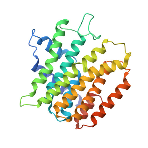 |
UniProt | |||||
Find proteins for Q6QLV2 (Antirrhinum majus) Explore Q6QLV2 Go to UniProtKB: Q6QLV2 | |||||
Entity Groups | |||||
| Sequence Clusters | 30% Identity50% Identity70% Identity90% Identity95% Identity100% Identity | ||||
| UniProt Group | Q6QLV2 | ||||
Sequence AnnotationsExpand | |||||
| |||||
| Length ( Å ) | Angle ( ˚ ) |
|---|---|
| a = 43.199 | α = 66.69 |
| b = 48.789 | β = 89.28 |
| c = 61.593 | γ = 89.98 |
| Software Name | Purpose |
|---|---|
| SCALEPACK | data scaling |
| PHASER | phasing |
| REFMAC | refinement |
| PDB_EXTRACT | data extraction |
| CBASS | data collection |
| HKL-2000 | data reduction |
| PHENIX | phasing |
RCSB PDB (citation) is hosted by
RCSB PDB is a member of the

