3QFV

MRCK beta in complex with TPCA-1


Experimental Data Snapshot

  • Method: X-RAY DIFFRACTION
  • Resolution: 2.65 Å
  • R-Value Free: 0.268 
  • R-Value Work: 0.192 
  • R-Value Observed: 0.196 

wwPDB Validation   3D Report Full Report


Ligand Structure Quality Assessment 


This is version 1.2 of the entry. See complete history


Literature

Co-crystal structures of inhibitors with MRCK beta , a key regulator of tumor cell invasion.

Heikkila, T.Wheatley, E.Crighton, D.Schroder, E.Boakes, A.Kaye, S.J.Mezna, M.Pang, L.Rushbrooke, M.Turnbull, A.Olson, M.F.

(2011) PLoS One 6: e24825-e24825

  • DOI: https://doi.org/10.1371/journal.pone.0024825
  • Primary Citation of Related Structures:  
    3QFV, 3TKU

  • PubMed Abstract: 

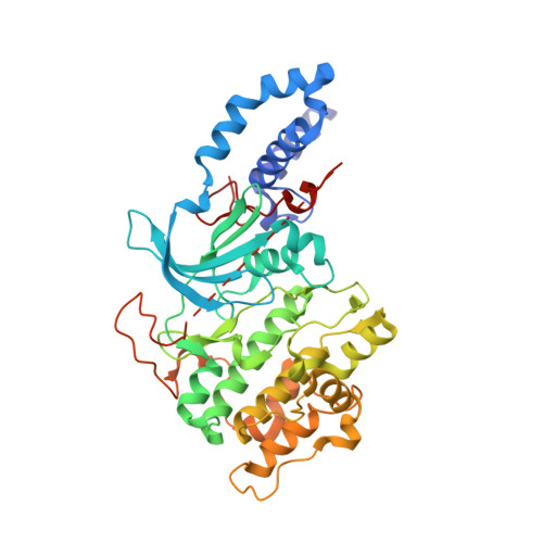
    MRCKα and MRCKβ (myotonic dystrophy kinase-related Cdc42-binding kinases) belong to a subfamily of Rho GTPase activated serine/threonine kinases within the AGC-family that regulate the actomyosin cytoskeleton. Reflecting their roles in myosin light chain (MLC) phosphorylation, MRCKα and MRCKβ influence cell shape and motility. We report further evidence for MRCKα and MRCKβ contributions to the invasion of cancer cells in 3-dimensional matrix invasion assays. In particular, our results indicate that the combined inhibition of MRCKα and MRCKβ together with inhibition of ROCK kinases results in significantly greater effects on reducing cancer cell invasion than blocking either MRCK or ROCK kinases alone. To probe the kinase ligand pocket, we screened 159 kinase inhibitors in an in vitro MRCKβ kinase assay and found 11 compounds that inhibited enzyme activity >80% at 3 µM. Further analysis of three hits, Y-27632, Fasudil and TPCA-1, revealed low micromolar IC(50) values for MRCKα and MRCKβ. We also describe the crystal structure of MRCKβ in complex with inhibitors Fasudil and TPCA-1 bound to the active site of the kinase. These high-resolution structures reveal a highly conserved AGC kinase fold in a typical dimeric arrangement. The kinase domain is in an active conformation with a fully-ordered and correctly positioned αC helix and catalytic residues in a conformation competent for catalysis. Together, these results provide further validation for MRCK involvement in regulation of cancer cell invasion and present a valuable starting point for future structure-based drug discovery efforts.


  • Organizational Affiliation

    Cancer Research Technology Discovery Laboratories, Wolfson Institute for Biomedical Research, London, United Kingdom.


Macromolecules
Find similar proteins by:  (by identity cutoff)  |  3D Structure
Entity ID: 1
MoleculeChains Sequence LengthOrganismDetailsImage
CDC42BPB protein
A, B
415Homo sapiensMutation(s): 0 
Gene Names: CDC42BPBMRCK beta
UniProt & NIH Common Fund Data Resources
Find proteins for Q9Y5S2 (Homo sapiens)
Explore Q9Y5S2 
Go to UniProtKB:  Q9Y5S2
PHAROS:  Q9Y5S2
GTEx:  ENSG00000198752 
Entity Groups  
Sequence Clusters30% Identity50% Identity70% Identity90% Identity95% Identity100% Identity
UniProt GroupQ9Y5S2
Sequence Annotations
Expand
  • Reference Sequence
Small Molecules
Ligands 4 Unique
IDChains Name / Formula / InChI Key2D Diagram3D Interactions
NM7
Query on NM7

Download Ideal Coordinates CCD File 
C [auth A],
E [auth B]
2-(carbamoylamino)-5-(4-fluorophenyl)thiophene-3-carboxamide
C12 H10 F N3 O2 S
SAYGKHKXGCPTLX-UHFFFAOYSA-N
EDO
Query on EDO

Download Ideal Coordinates CCD File 
D [auth A],
I [auth B],
J [auth B]
1,2-ETHANEDIOL
C2 H6 O2
LYCAIKOWRPUZTN-UHFFFAOYSA-N
CL
Query on CL

Download Ideal Coordinates CCD File 
F [auth B],
G [auth B]
CHLORIDE ION
Cl
VEXZGXHMUGYJMC-UHFFFAOYSA-M
MG
Query on MG

Download Ideal Coordinates CCD File 
H [auth B]MAGNESIUM ION
Mg
JLVVSXFLKOJNIY-UHFFFAOYSA-N
Binding Affinity Annotations 
IDSourceBinding Affinity
NM7 PDBBind:  3QFV IC50: 1690 (nM) from 1 assay(s)
Experimental Data & Validation

Experimental Data

  • Method: X-RAY DIFFRACTION
  • Resolution: 2.65 Å
  • R-Value Free: 0.268 
  • R-Value Work: 0.192 
  • R-Value Observed: 0.196 
  • Space Group: P 1 21 1
Unit Cell:
Length ( Å )Angle ( ˚ )
a = 44.81α = 90
b = 123.36β = 100.69
c = 85.49γ = 90
Software Package:
Software NamePurpose
DNAdata collection
PHASERphasing
REFMACrefinement
MOSFLMdata reduction
SCALAdata scaling

Structure Validation

View Full Validation Report


Ligand Structure Quality Assessment 


Entry History 

Deposition Data

Revision History  (Full details and data files)

  • Version 1.0: 2011-10-12
    Type: Initial release
  • Version 1.1: 2013-08-28
    Changes: Database references
  • Version 1.2: 2024-02-21
    Changes: Data collection, Database references