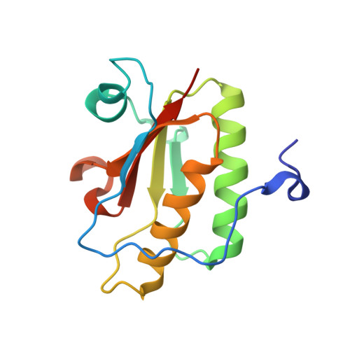
3QD7

Crystal structure of YdaL, a stand-alone small MutS-related protein from Escherichia coli


Experimental Data Snapshot

  • Method: X-RAY DIFFRACTION
  • Resolution: 2.30 Å
  • R-Value Free: 0.244 
  • R-Value Work: 0.224 
  • R-Value Observed: 0.225 

wwPDB Validation   3D Report Full Report


This is version 1.2 of the entry. See complete history


Literature

Crystal structure of YdaL, a stand-alone small MutS-related protein from Escherichia coli.

Gui, W.J.Qu, Q.H.Chen, Y.Y.Wang, M.Zhang, X.E.Bi, L.J.Jiang, T.

(2011) J Struct Biol 174: 282-289

  • DOI: https://doi.org/10.1016/j.jsb.2011.01.008
  • Primary Citation of Related Structures:  
    3QD7

  • PubMed Abstract: 

    Sequence homologs of the small MutS-related (Smr) domain, the C-terminal endonuclease domain of MutS2, also exist as stand-alone proteins. In this study, we report the crystal structure of a proteolyzed fragment of YdaL (YdaL₃₉-₁₇₅), a stand-alone Smr protein from Escherichia coli. In this structure, residues 86-170 assemble into a classical Smr core domain and are embraced by an N-terminal extension (residues 40-85) with an α/β/α fold. Sequence alignment indicates that the N-terminal extension is conserved among a number of stand-alone Smr proteins, suggesting structural diversity among Smr domains. We also discovered that the DNA binding affinity and endonuclease activity of the truncated YdaL₃₉-₁₇₅ protein were slightly lower than those of full-length YdaL₁-₁₈₇, suggesting that residues 1-38 may be involved in DNA binding.


  • Organizational Affiliation

    National Laboratory of Biomacromolecules, Institute of Biophysics, Chinese Academy of Sciences, 15 Datun Road, Chaoyang District, Beijing 100101, China.


Macromolecules
Find similar proteins by:  (by identity cutoff)  |  3D Structure
Entity ID: 1
MoleculeChains Sequence LengthOrganismDetailsImage
Uncharacterized protein ydaLA [auth X]137Escherichia coli K-12Mutation(s): 0 
Gene Names: ydaLb1340JW1334
UniProt
Find proteins for P76053 (Escherichia coli (strain K12))
Explore P76053 
Go to UniProtKB:  P76053
Entity Groups  
Sequence Clusters30% Identity50% Identity70% Identity90% Identity95% Identity100% Identity
UniProt GroupP76053
Sequence Annotations
Expand
  • Reference Sequence
Experimental Data & Validation

Experimental Data

  • Method: X-RAY DIFFRACTION
  • Resolution: 2.30 Å
  • R-Value Free: 0.244 
  • R-Value Work: 0.224 
  • R-Value Observed: 0.225 
  • Space Group: P 21 21 2
Unit Cell:
Length ( Å )Angle ( ˚ )
a = 95.517α = 90
b = 34.833β = 90
c = 40.698γ = 90
Software Package:
Software NamePurpose
CrystalCleardata collection
PHENIXmodel building
REFMACrefinement
HKL-2000data reduction
SCALAdata scaling
PHENIXphasing

Structure Validation

View Full Validation Report


Entry History 

Deposition Data

Revision History  (Full details and data files)

  • Version 1.0: 2011-06-22
    Type: Initial release
  • Version 1.1: 2011-07-13
    Changes: Version format compliance
  • Version 1.2: 2024-03-20
    Changes: Data collection, Database references