


Crystal structure of the 3-Dehydroquinate Synthase (aroB) from Mycobacterium tuberculosis
Cheng, W.C., Chen, T.J., Wang, W.C.To be published.
Experimental Data Snapshot
wwPDB Validation 3D Report Full Report

Entity ID: 1 | |||||
|---|---|---|---|---|---|
| Molecule | Chains | Sequence Length | Organism | Details | Image |
| 3-dehydroquinate synthase | 368 | Mycobacterium tuberculosis H37Rv | Mutation(s): 0 Gene Names: aroB, MRA_2566 EC: 4.2.3.4 | 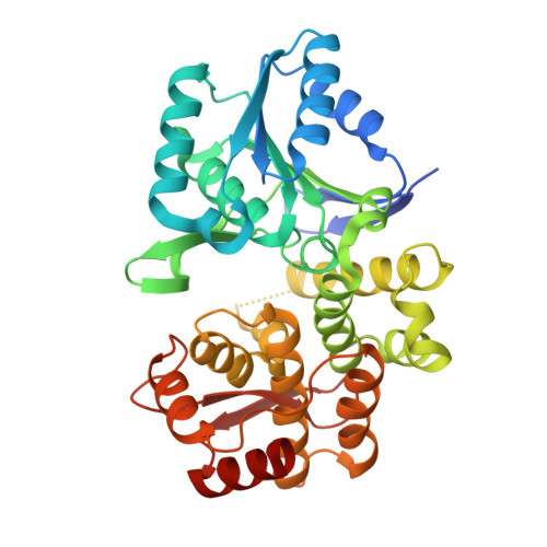 | |
UniProt | |||||
Find proteins for P9WPX9 (Mycobacterium tuberculosis (strain ATCC 25618 / H37Rv)) Explore P9WPX9 Go to UniProtKB: P9WPX9 | |||||
Entity Groups | |||||
| Sequence Clusters | 30% Identity50% Identity70% Identity90% Identity95% Identity100% Identity | ||||
| UniProt Group | P9WPX9 | ||||
Sequence AnnotationsExpand | |||||
| |||||
| Ligands 2 Unique | |||||
|---|---|---|---|---|---|
| ID | Chains | Name / Formula / InChI Key | 2D Diagram | 3D Interactions | |
| ZN Query on ZN | B [auth A], C [auth A] | ZINC ION Zn PTFCDOFLOPIGGS-UHFFFAOYSA-N |  | ||
| CL Query on CL | D [auth A] | CHLORIDE ION Cl VEXZGXHMUGYJMC-UHFFFAOYSA-M |  | ||
| Length ( Å ) | Angle ( ˚ ) |
|---|---|
| a = 140.318 | α = 90 |
| b = 140.318 | β = 90 |
| c = 38.408 | γ = 90 |
| Software Name | Purpose |
|---|---|
| HKL-2000 | data collection |
| AMoRE | phasing |
| REFMAC | refinement |
| HKL-2000 | data reduction |
| HKL-2000 | data scaling |
RCSB PDB (citation) is hosted by
RCSB PDB is a member of the

