3Q7E

Crystal Structure of rat Protein Arginine Methyltransferase 1 (PRMT1)M48L mutant


Experimental Data Snapshot

  • Method: X-RAY DIFFRACTION
  • Resolution: 2.20 Å
  • R-Value Free: 0.245 
  • R-Value Work: 0.201 
  • R-Value Observed: 0.203 

wwPDB Validation   3D Report Full Report


Ligand Structure Quality Assessment 


This is version 1.3 of the entry. See complete history


Literature

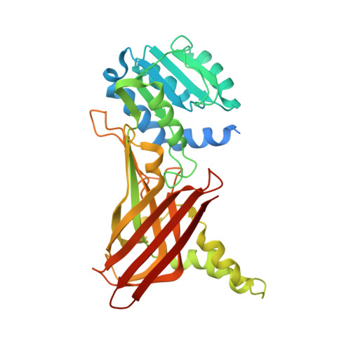
Investigation of the molecular origins of protein-arginine methyltransferase I (PRMT1) product specificity reveals a role for two conserved methionine residues.

Gui, S.Wooderchak, W.L.Daly, M.P.Porter, P.J.Johnson, S.J.Hevel, J.M.

(2011) J Biol Chem 286: 29118-29126

  • DOI: https://doi.org/10.1074/jbc.M111.224097
  • Primary Citation of Related Structures:  
    3Q7E

  • PubMed Abstract: 

    Protein-arginine methyltransferases aid in the regulation of many biological processes by methylating specific arginyl groups within targeted proteins. The varied nature of the response to methylation is due in part to the diverse product specificity displayed by the protein-arginine methyltransferases. In addition to site location within a protein, biological response is also determined by the degree (mono-/dimethylation) and type of arginine dimethylation (asymmetric/symmetric). Here, we have identified two strictly conserved methionine residues in the PRMT1 active site that are not only important for activity but also control substrate specificity. Mutation of Met-155 or Met-48 results in a loss in activity and a change in distribution of mono- and dimethylated products. The altered substrate specificity of M155A and M48L mutants is also evidenced by automethylation. Investigation into the mechanistic basis of altered substrate recognition led us to consider each methyl transfer step separately. Single turnover experiments reveal that the rate of transfer of the second methyl group is much slower than transfer of the first methyl group in M48L, especially for arginine residues located in the center of the peptide substrate where turnover of the monomethylated species is negligible. Thus, altered product specificity in M48L originates from the differential effect of the mutation on the two rates. Characterization of the two active-site methionines provides the first insight into how the PRMT1 active site is engineered to control product specificity.


  • Organizational Affiliation

    Chemistry and Biochemistry Department, Utah State University, Logan, Utah 84322 and.


Macromolecules
Find similar proteins by:  (by identity cutoff)  |  3D Structure
Entity ID: 1
MoleculeChains Sequence LengthOrganismDetailsImage
Protein arginine N-methyltransferase 1349Rattus norvegicusMutation(s): 1 
Gene Names: Prmt1Hrmt1l2
EC: 2.1.1
UniProt
Find proteins for Q63009 (Rattus norvegicus)
Explore Q63009 
Go to UniProtKB:  Q63009
Entity Groups  
Sequence Clusters30% Identity50% Identity70% Identity90% Identity95% Identity100% Identity
UniProt GroupQ63009
Sequence Annotations
Expand
  • Reference Sequence
Small Molecules
Ligands 2 Unique
IDChains Name / Formula / InChI Key2D Diagram3D Interactions
SAH
Query on SAH

Download Ideal Coordinates CCD File 
B [auth A]S-ADENOSYL-L-HOMOCYSTEINE
C14 H20 N6 O5 S
ZJUKTBDSGOFHSH-WFMPWKQPSA-N
MG
Query on MG

Download Ideal Coordinates CCD File 
C [auth A]MAGNESIUM ION
Mg
JLVVSXFLKOJNIY-UHFFFAOYSA-N
Binding Affinity Annotations 
IDSourceBinding Affinity
SAH BindingDB:  3Q7E Ki: 400 (nM) from 1 assay(s)
IC50: min: 420, max: 550 (nM) from 3 assay(s)
Experimental Data & Validation

Experimental Data

  • Method: X-RAY DIFFRACTION
  • Resolution: 2.20 Å
  • R-Value Free: 0.245 
  • R-Value Work: 0.201 
  • R-Value Observed: 0.203 
  • Space Group: P 41 2 2
Unit Cell:
Length ( Å )Angle ( ˚ )
a = 87.307α = 90
b = 87.307β = 90
c = 143.29γ = 90
Software Package:
Software NamePurpose
d*TREKdata scaling
PHASERphasing
PHENIXrefinement
PDB_EXTRACTdata extraction
CrystalCleardata collection
d*TREKdata reduction

Structure Validation

View Full Validation Report


Ligand Structure Quality Assessment 


Entry History 

Deposition Data

Revision History  (Full details and data files)

  • Version 1.0: 2011-06-22
    Type: Initial release
  • Version 1.1: 2011-07-13
    Changes: Version format compliance
  • Version 1.2: 2013-01-09
    Changes: Database references
  • Version 1.3: 2023-09-13
    Changes: Data collection, Database references, Derived calculations, Refinement description