3Q3D

Crystal structure of BmrR bound to puromycin


Experimental Data Snapshot

  • Method: X-RAY DIFFRACTION
  • Resolution: 2.79 Å
  • R-Value Free: 0.262 
  • R-Value Work: 0.218 
  • R-Value Observed: 0.220 

wwPDB Validation   3D Report Full Report


Ligand Structure Quality Assessment 


This is version 1.5 of the entry. See complete history


Literature

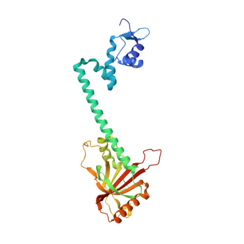
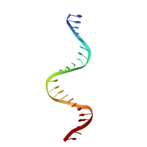
Structural contributions to multidrug recognition in the multidrug resistance (MDR) gene regulator, BmrR.

Bachas, S.Eginton, C.Gunio, D.Wade, H.

(2011) Proc Natl Acad Sci U S A 108: 11046-11051

  • DOI: https://doi.org/10.1073/pnas.1104850108
  • Primary Citation of Related Structures:  
    3Q1M, 3Q2Y, 3Q3D, 3Q5P, 3Q5R, 3Q5S

  • PubMed Abstract: 

    Current views of multidrug (MD) recognition focus on large drug-binding cavities with flexible elements. However, MD recognition in BmrR is supported by a small, rigid drug-binding pocket. Here, a detailed description of MD binding by the noncanonical BmrR protein is offered through the combined use of X-ray and solution studies. Low shape complementarity, suboptimal packing, and efficient burial of a diverse set of ligands is facilitated by an aromatic docking platform formed by a set of conformationally fixed aromatic residues, hydrophobic pincer pair that locks the different drug structures on the adaptable platform surface, and a trio of acidic residues that enables cation selectivity without much regard to ligand structure. Within the binding pocket is a set of BmrR-derived H-bonding donor and acceptors that solvate a wide range of ligand polar substituent arrangements in a manner analogous to aqueous solvent. Energetic analyses of MD binding by BmrR are consistent with structural data. A common binding orientation for the different BmrR ligands is in line with promiscuous allosteric regulation.


  • Organizational Affiliation

    Department of Biophysics and Biophysical Chemistry, Johns Hopkins School of Medicine, Baltimore, MD 21205, USA.


Macromolecules

Find similar proteins by:  (by identity cutoff)  |  3D Structure
Entity ID: 1
MoleculeChains Sequence LengthOrganismDetailsImage
Multidrug-efflux transporter 1 regulator284Bacillus subtilisMutation(s): 0 
Gene Names: bmrRbmr1RBSU24020
UniProt
Find proteins for P39075 (Bacillus subtilis (strain 168))
Explore P39075 
Go to UniProtKB:  P39075
Entity Groups  
Sequence Clusters30% Identity50% Identity70% Identity90% Identity95% Identity100% Identity
UniProt GroupP39075
Sequence Annotations
Expand
  • Reference Sequence

Find similar nucleic acids by:  Sequence   |   3D Structure  

Entity ID: 2
MoleculeChains LengthOrganismImage
BmrR promoter DNA23N/A
Sequence Annotations
Expand
  • Reference Sequence
Small Molecules
Ligands 1 Unique
IDChains Name / Formula / InChI Key2D Diagram3D Interactions
PUY
Query on PUY

Download Ideal Coordinates CCD File 
C [auth A]PUROMYCIN
C22 H29 N7 O5
RXWNCPJZOCPEPQ-NVWDDTSBSA-N
Experimental Data & Validation

Experimental Data

  • Method: X-RAY DIFFRACTION
  • Resolution: 2.79 Å
  • R-Value Free: 0.262 
  • R-Value Work: 0.218 
  • R-Value Observed: 0.220 
  • Space Group: P 43 2 2
Unit Cell:
Length ( Å )Angle ( ˚ )
a = 106.486α = 90
b = 106.486β = 90
c = 145.403γ = 90
Software Package:
Software NamePurpose
REFMACrefinement
CNSrefinement
MAR345dtbdata collection
HKL-2000data reduction
HKL-2000data scaling
CNSphasing

Structure Validation

View Full Validation Report


Ligand Structure Quality Assessment 


Entry History 

Deposition Data

Revision History  (Full details and data files)

  • Version 1.0: 2011-06-15
    Type: Initial release
  • Version 1.1: 2011-07-13
    Changes: Version format compliance
  • Version 1.2: 2011-07-20
    Changes: Database references
  • Version 1.3: 2019-07-17
    Changes: Data collection, Refinement description
  • Version 1.4: 2024-02-21
    Changes: Data collection, Database references, Derived calculations
  • Version 1.5: 2024-03-13
    Changes: Source and taxonomy