


Crystal Structure of Dihydrofolate Reductase from Yersinia pestis
Maltseva, N., Kim, Y., Makowska-Grzyska, M., Mulligan, R., Papazisi, L., Anderson, W.F., Joachimiak, A., Center for Structural Genomics of Infectious Diseases (CSGID)To be published.
Experimental Data Snapshot
wwPDB Validation 3D Report Full Report

Entity ID: 1 | |||||
|---|---|---|---|---|---|
| Molecule | Chains | Sequence Length | Organism | Details | Image |
| Dihydrofolate reductase | 163 | Yersinia pestis CO92 | Mutation(s): 0 Gene Names: folA, y3688, YPO0486, YP_3693 EC: 1.5.1.3 | 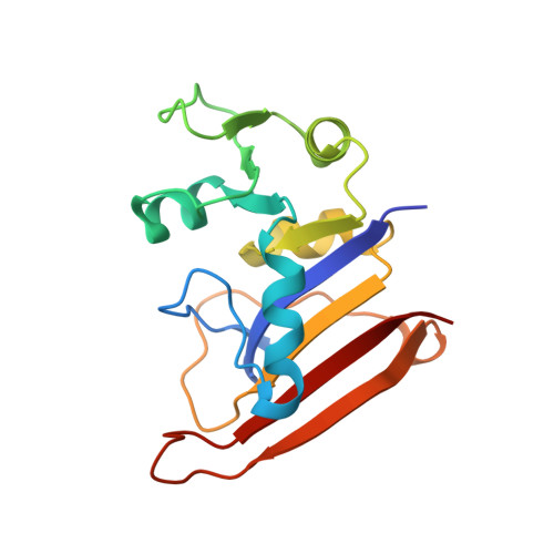 | |
UniProt | |||||
Find proteins for A0A3N4BLI0 (Yersinia pestis) Explore A0A3N4BLI0 Go to UniProtKB: A0A3N4BLI0 | |||||
Entity Groups | |||||
| Sequence Clusters | 30% Identity50% Identity70% Identity90% Identity95% Identity100% Identity | ||||
| UniProt Group | A0A3N4BLI0 | ||||
Sequence AnnotationsExpand | |||||
| |||||
| Ligands 1 Unique | |||||
|---|---|---|---|---|---|
| ID | Chains | Name / Formula / InChI Key | 2D Diagram | 3D Interactions | |
| SO4 Query on SO4 | B [auth A], C [auth A] | SULFATE ION O4 S QAOWNCQODCNURD-UHFFFAOYSA-L |  | ||
| Length ( Å ) | Angle ( ˚ ) |
|---|---|
| a = 60.477 | α = 90 |
| b = 79.152 | β = 90 |
| c = 64.685 | γ = 90 |
| Software Name | Purpose |
|---|---|
| SBC-Collect | data collection |
| HKL-3000 | data collection |
| BALBES | phasing |
| PHENIX | refinement |
| HKL-3000 | data reduction |
| HKL-3000 | data scaling |
RCSB PDB (citation) is hosted by
RCSB PDB is a member of the

