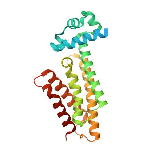
Structural activation of the transcriptional repressor EthR from Mycobacterium tuberculosis by single amino acid change mimicking natural and synthetic ligands.
Carette, X., Blondiaux, N., Willery, E., Hoos, S., Lecat-Guillet, N., Lens, Z., Wohlkonig, A., Wintjens, R., Soror, S.H., Frenois, F., Dirie, B., Villeret, V., England, P., Lippens, G., Deprez, B., Locht, C., Willand, N., Baulard, A.R.(2012) Nucleic Acids Res 40: 3018-3030
- PubMed: 22156370
- DOI: https://doi.org/10.1093/nar/gkr1113
- Primary Citation of Related Structures:
3Q0W, 3QPL, 3TP0, 3TP3 - PubMed Abstract:
Ethionamide is an antituberculous drug for the treatment of multidrug-resistant Mycobacterium tuberculosis. This antibiotic requires activation by the monooxygenase EthA to exert its activity. Production of EthA is controlled by the transcriptional repressor EthR, a member of the TetR family. The sensitivity of M. tuberculosis to ethionamide can be artificially enhanced using synthetic ligands of EthR that allosterically inactivate its DNA-binding activity. Comparison of several structures of EthR co-crystallized with various ligands suggested that the structural reorganization of EthR resulting in its inactivation is controlled by a limited portion of the ligand-binding-pocket. In silico simulation predicted that mutation G106W may mimic ligands. X-ray crystallography of variant G106W indeed revealed a protein structurally similar to ligand-bound EthR. Surface plasmon resonance experiments established that this variant is unable to bind DNA, while thermal shift studies demonstrated that mutation G106W stabilizes EthR as strongly as ligands. Proton NMR of the methyl regions showed a lesser contribution of exchange broadening upon ligand binding, and the same quenched dynamics was observed in apo-variant G106W. Altogether, we here show that the area surrounding Gly106 constitutes the molecular switch involved in the conformational reorganization of EthR. These results also shed light on the mechanistic of ligand-induced allosterism controlling the DNA binding properties of TetR family repressors.
Organizational Affiliation:
Center for Infection and Immunity of Lille, F-59019 Lille, France.