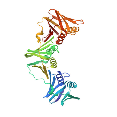
3PWE

Crystal structure of the E. coli beta clamp mutant R103C, I305C, C260S, C333S at 2.2A resolution


Experimental Data Snapshot

  • Method: X-RAY DIFFRACTION
  • Resolution: 2.20 Å
  • R-Value Free: 0.245 
  • R-Value Work: 0.204 
  • R-Value Observed: 0.206 

wwPDB Validation   3D Report Full Report


This is version 1.4 of the entry. See complete history


Literature

The E. coli clamp loader can actively pry open the beta-sliding clamp

Paschall, C.O.Thompson, J.A.Marzahn, M.R.Chiraniya, A.Hayner, J.N.O'Donnell, M.Robbins, A.H.McKenna, R.Bloom, L.B.

(2011) J Biol Chem 286: 42704-42714

  • DOI: https://doi.org/10.1074/jbc.M111.268169
  • Primary Citation of Related Structures:  
    3PWE

  • PubMed Abstract: 

    Clamp loaders load ring-shaped sliding clamps onto DNA. Once loaded onto DNA, sliding clamps bind to DNA polymerases to increase the processivity of DNA synthesis. To load clamps onto DNA, an open clamp loader-clamp complex must form. An unresolved question is whether clamp loaders capture clamps that have transiently opened or whether clamp loaders bind closed clamps and actively open clamps. A simple fluorescence-based clamp opening assay was developed to address this question and to determine how ATP binding contributes to clamp opening. A direct comparison of real time binding and opening reactions revealed that the Escherichia coli γ complex binds β first and then opens the clamp. Mutation of conserved "arginine fingers" in the γ complex that interact with bound ATP decreased clamp opening activity showing that arginine fingers make an important contribution to the ATP-induced conformational changes that allow the clamp loader to pry open the clamp.


  • Organizational Affiliation

    Department of Biochemistry and Molecular Biology, University of Florida, Gainesville, Florida 32610-0245.


Macromolecules
Find similar proteins by:  (by identity cutoff)  |  3D Structure
Entity ID: 1
MoleculeChains Sequence LengthOrganismDetailsImage
DNA polymerase III subunit beta
A, B
366Escherichia coli K-12Mutation(s): 4 
Gene Names: dnaNb3701JW3678
EC: 2.7.7.7
UniProt
Find proteins for P0A988 (Escherichia coli (strain K12))
Explore P0A988 
Go to UniProtKB:  P0A988
Entity Groups  
Sequence Clusters30% Identity50% Identity70% Identity90% Identity95% Identity100% Identity
UniProt GroupP0A988
Sequence Annotations
Expand
  • Reference Sequence
Experimental Data & Validation

Experimental Data

  • Method: X-RAY DIFFRACTION
  • Resolution: 2.20 Å
  • R-Value Free: 0.245 
  • R-Value Work: 0.204 
  • R-Value Observed: 0.206 
  • Space Group: P 1 21 1
Unit Cell:
Length ( Å )Angle ( ˚ )
a = 79.799α = 90
b = 67.362β = 114.16
c = 80.742γ = 90
Software Package:
Software NamePurpose
DENZOdata reduction
SCALEPACKdata scaling
PHENIXrefinement
PDB_EXTRACTdata extraction
CrystalCleardata collection
HKL-2000data reduction
HKL-2000data scaling
PHENIXphasing

Structure Validation

View Full Validation Report


Entry History 

Deposition Data

Revision History  (Full details and data files)

  • Version 1.0: 2011-10-19
    Type: Initial release
  • Version 1.1: 2011-11-23
    Changes: Database references
  • Version 1.2: 2013-06-26
    Changes: Database references
  • Version 1.3: 2017-11-08
    Changes: Refinement description
  • Version 1.4: 2023-09-13
    Changes: Data collection, Database references, Refinement description