3PVF

Structure of C126S mutant of Plasmodium falciparum triosephosphate isomerase complexed with PGA


Experimental Data Snapshot

  • Method: X-RAY DIFFRACTION
  • Resolution: 1.73 Å
  • R-Value Free: 0.194 
  • R-Value Work: 0.152 
  • R-Value Observed: 0.154 

wwPDB Validation   3D Report Full Report


Ligand Structure Quality Assessment 


This is version 1.3 of the entry. See complete history


Literature

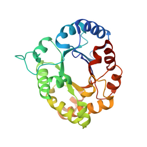
Probing the role of the fully conserved Cys126 in triosephosphate isomerase by site-specific mutagenesis--distal effects on dimer stability.

Samanta, M.Banerjee, M.Murthy, M.R.Balaram, H.Balaram, P.

(2011) FEBS J 278: 1932-1943

  • DOI: https://doi.org/10.1111/j.1742-4658.2011.08110.x
  • Primary Citation of Related Structures:  
    3PVF, 3PWA, 3PY2

  • PubMed Abstract: 

    Cys126 is a completely conserved residue in triosephosphate isomerase that is proximal to the active site but has been ascribed no specific role in catalysis. A previous study of the C126S and C126A mutants of yeast TIM reported substantial catalytic activity for the mutant enzymes, leading to the suggestion that this residue is implicated in folding and stability [Gonzalez-Mondragon E et al. (2004) Biochemistry 43, 3255-3263]. We re-examined the role of Cys126 with the Plasmodium falciparum enzyme as a model. Five mutants, C126S, C126A, C126V, C126M, and C126T, were characterized. Crystal structures of the 3-phosphoglycolate-bound C126S mutant and the unliganded forms of the C126S and C126A mutants were determined at a resolution of 1.7-2.1 Å. Kinetic studies revealed an approximately five-fold drop in k(cat) for the C126S and C126A mutants, whereas an approximately 10-fold drop was observed for the other three mutants. At ambient temperature, the wild-type enzyme and all five mutants showed no concentration dependence of activity. At higher temperatures (> 40 °C), the mutants showed a significant concentration dependence, with a dramatic loss in activity below 15 μM. The mutants also had diminished thermal stability at low concentration, as monitored by far-UV CD. These results suggest that Cys126 contributes to the stability of the dimer interface through a network of interactions involving His95, Glu97, and Arg98, which form direct contacts across the dimer interface.


  • Organizational Affiliation

    Molecular Biophysics Unit, Indian Institute of Science, Bangalore, India.


Macromolecules
Find similar proteins by:  (by identity cutoff)  |  3D Structure
Entity ID: 1
MoleculeChains Sequence LengthOrganismDetailsImage
Triosephosphate isomerase248Plasmodium falciparumMutation(s): 1 
Gene Names: TPI
EC: 5.3.1.1
UniProt
Find proteins for Q07412 (Plasmodium falciparum)
Explore Q07412 
Go to UniProtKB:  Q07412
Entity Groups  
Sequence Clusters30% Identity50% Identity70% Identity90% Identity95% Identity100% Identity
UniProt GroupQ07412
Sequence Annotations
Expand
  • Reference Sequence
Small Molecules
Ligands 1 Unique
IDChains Name / Formula / InChI Key2D Diagram3D Interactions
PGA
Query on PGA

Download Ideal Coordinates CCD File 
B [auth A]2-PHOSPHOGLYCOLIC ACID
C2 H5 O6 P
ASCFNMCAHFUBCO-UHFFFAOYSA-N
Experimental Data & Validation

Experimental Data

  • Method: X-RAY DIFFRACTION
  • Resolution: 1.73 Å
  • R-Value Free: 0.194 
  • R-Value Work: 0.152 
  • R-Value Observed: 0.154 
  • Space Group: C 1 2 1
Unit Cell:
Length ( Å )Angle ( ˚ )
a = 87.51α = 90
b = 63.1β = 117.23
c = 53.2γ = 90
Software Package:
Software NamePurpose
MAR345dtbdata collection
PHASERphasing
REFMACrefinement
MOSFLMdata reduction
SCALAdata scaling

Structure Validation

View Full Validation Report


Ligand Structure Quality Assessment 


Entry History 

Deposition Data

Revision History  (Full details and data files)

  • Version 1.0: 2011-04-27
    Type: Initial release
  • Version 1.1: 2011-07-13
    Changes: Version format compliance
  • Version 1.2: 2013-08-28
    Changes: Database references
  • Version 1.3: 2023-11-01
    Changes: Data collection, Database references, Derived calculations, Refinement description