3PT3

Crystal structure of the C-terminal lobe of the human UBR5 HECT domain


Experimental Data Snapshot

  • Method: X-RAY DIFFRACTION
  • Resolution: 1.97 Å
  • R-Value Free: 0.276 
  • R-Value Work: 0.219 
  • R-Value Observed: 0.222 

wwPDB Validation   3D Report Full Report


This is version 1.2 of the entry. See complete history


Literature

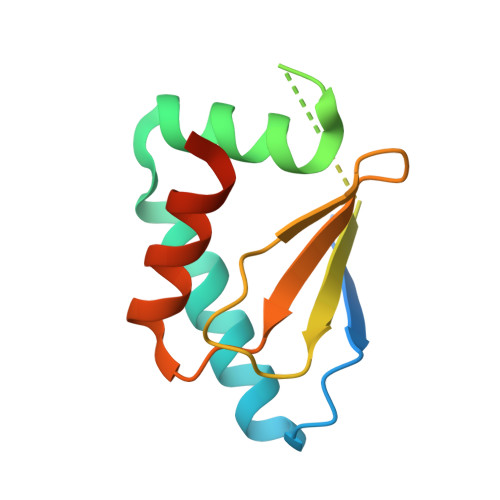
Structure of the HECT C-lobe of the UBR5 E3 ubiquitin ligase.

Matta-Camacho, E.Kozlov, G.Menade, M.Gehring, K.

(2012) Acta Crystallogr Sect F Struct Biol Cryst Commun 68: 1158-1163

  • DOI: https://doi.org/10.1107/S1744309112036937
  • Primary Citation of Related Structures:  
    3PT3

  • PubMed Abstract: 

    UBR5 ubiquitin ligase (also known as EDD, Rat100 or hHYD) is a member of the E3 protein family of HECT (homologous to E6-AP C-terminus) ligases as it contains a C-terminal HECT domain. In ubiquitination cascades involving E3s of the HECT class, ubiquitin is transferred from an associated E2 ubiquitin-conjugating enzyme to the acceptor cysteine of the HECT domain, which consists of structurally distinct N- and C-lobes connected by a flexible linker. Here, the high-resolution crystal structure of the C-lobe of the HECT domain of human UBR5 is presented. The structure reveals important features that are unique compared with other HECT domains. In particular, a distinct four-residue insert in the second helix elongates this helix, resulting in a strikingly different orientation of the preceding loop. This protruding loop is likely to contribute to specificity towards the E2 ubiquitin-conjugating enzyme UBCH4, which is an important functional partner of UBR5. Ubiquitination assays showed that the C-lobe of UBR5 is able to form a thioester-linked E3-ubiquitin complex, although it does not physically interact with UBCH4 in NMR experiments. This study contributes to a better understanding of UBR5 ubiquitination activity.


  • Organizational Affiliation

    Groupe de Recherche axé sur la Structure des Protéines, Department of Biochemistry, McGill University, Montreal, Quebec H3G 0B1, Canada.


Macromolecules
Find similar proteins by:  (by identity cutoff)  |  3D Structure
Entity ID: 1
MoleculeChains Sequence LengthOrganismDetailsImage
E3 ubiquitin-protein ligase UBR5
A, B
118Homo sapiensMutation(s): 0 
Gene Names: UBR5EDDEDD1HYDKIAA0896
EC: 6.3.2
UniProt & NIH Common Fund Data Resources
Find proteins for O95071 (Homo sapiens)
Explore O95071 
Go to UniProtKB:  O95071
PHAROS:  O95071
GTEx:  ENSG00000104517 
Entity Groups  
Sequence Clusters30% Identity50% Identity70% Identity90% Identity95% Identity100% Identity
UniProt GroupO95071
Sequence Annotations
Expand
  • Reference Sequence
Experimental Data & Validation

Experimental Data

  • Method: X-RAY DIFFRACTION
  • Resolution: 1.97 Å
  • R-Value Free: 0.276 
  • R-Value Work: 0.219 
  • R-Value Observed: 0.222 
  • Space Group: P 1
Unit Cell:
Length ( Å )Angle ( ˚ )
a = 29.24α = 90.04
b = 39.07β = 84.59
c = 44.17γ = 81.77
Software Package:
Software NamePurpose
ADSCdata collection
PHASERphasing
REFMACrefinement
HKL-2000data reduction
HKL-2000data scaling

Structure Validation

View Full Validation Report


Entry History 

Deposition Data

Revision History  (Full details and data files)

  • Version 1.0: 2012-01-25
    Type: Initial release
  • Version 1.1: 2012-10-17
    Changes: Database references
  • Version 1.2: 2023-09-06
    Changes: Data collection, Database references, Refinement description