3PR5

Dpo4 Y12A mutant incorporating ADP opposite template dT


Experimental Data Snapshot

  • Method: X-RAY DIFFRACTION
  • Resolution: 2.40 Å
  • R-Value Free: 0.265 
  • R-Value Work: 0.228 
  • R-Value Observed: 0.229 

wwPDB Validation   3D Report Full Report


Ligand Structure Quality Assessment 


This is version 1.2 of the entry. See complete history


Literature

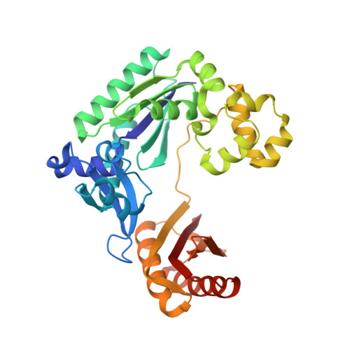
Structural mechanism of ribonucleotide discrimination by a Y-family DNA polymerase.

Kirouac, K.N.Suo, Z.Ling, H.

(2011) J Mol Biol 407: 382-390

  • DOI: https://doi.org/10.1016/j.jmb.2011.01.037
  • Primary Citation of Related Structures:  
    3PR4, 3PR5

  • PubMed Abstract: 

    The ability of DNA polymerases to differentiate between ribonucleotides and deoxribonucleotides is fundamental to the accurate replication and maintenance of an organism's genome. The active sites of Y-family DNA polymerases are highly solvent accessible, yet these enzymes still maintain a high selectivity towards deoxyribonucleotides. Here, we biochemically demonstrate that a single active-site mutation (Y12A) in Dpo4, a model Y-family DNA polymerase, causes both a dramatic loss of ribonucleotide discrimination and a decrease in nucleotide incorporation efficiency. We also determined two ternary crystal structures of the Dpo4 Y12A mutant incorporating either dATP or ATP nucleotides opposite a template dT base. Interestingly, both dATP and ATP were hydrolyzed to dADP and ADP, respectively. In addition, the dADP and ADP molecules adopt a similar conformation and position at the polymerase active site to a ddADP molecule in the ternary crystal structure of wild-type Dpo4. The Y12A mutant loses stacking interactions with the deoxyribose of dNTP, which destabilizes the binding of incoming nucleotides. The mutation also opens a space to accommodate the 2'-OH group of the ribose of NTP in the polymerase active site. The structural change leads to the reduction in deoxynucleotide incorporation efficiency and allows ribonucleotide incorporation.


  • Organizational Affiliation

    Department of Biochemistry, University of Western Ontario, London, Ontario, Canada N6A 5C1.


Macromolecules

Find similar proteins by:  (by identity cutoff)  |  3D Structure
Entity ID: 1
MoleculeChains Sequence LengthOrganismDetailsImage
DNA polymerase IVA [auth B]341Saccharolobus solfataricusMutation(s): 1 
Gene Names: dbhdpo4SSO2448
EC: 2.7.7.7
UniProt
Find proteins for Q97W02 (Saccharolobus solfataricus (strain ATCC 35092 / DSM 1617 / JCM 11322 / P2))
Explore Q97W02 
Go to UniProtKB:  Q97W02
Entity Groups  
Sequence Clusters30% Identity50% Identity70% Identity90% Identity95% Identity100% Identity
UniProt GroupQ97W02
Sequence Annotations
Expand
  • Reference Sequence

Find similar nucleic acids by:  Sequence   |   3D Structure  

Entity ID: 2
MoleculeChains LengthOrganismImage
DNA (5'-D(*GP*GP*GP*GP*GP*AP*AP*GP*GP*AP*CP*TP*C)-3')B [auth P]13N/A
Sequence Annotations
Expand
  • Reference Sequence

Find similar nucleic acids by:  Sequence   |   3D Structure  

Entity ID: 3
MoleculeChains LengthOrganismImage
DNA (5'-D(*TP*TP*CP*AP*TP*GP*AP*GP*TP*CP*CP*TP*TP*CP*CP*CP*CP*C)-3')C [auth T]18N/A
Sequence Annotations
Expand
  • Reference Sequence
Experimental Data & Validation

Experimental Data

  • Method: X-RAY DIFFRACTION
  • Resolution: 2.40 Å
  • R-Value Free: 0.265 
  • R-Value Work: 0.228 
  • R-Value Observed: 0.229 
  • Space Group: P 21 21 2
Unit Cell:
Length ( Å )Angle ( ˚ )
a = 95.607α = 90
b = 101.993β = 90
c = 52.288γ = 90
Software Package:
Software NamePurpose
MAR345dtbdata collection
PHASERphasing
PHENIXrefinement
DENZOdata reduction
SCALEPACKdata scaling

Structure Validation

View Full Validation Report


Ligand Structure Quality Assessment 


Entry History 

Deposition Data

Revision History  (Full details and data files)

  • Version 1.0: 2011-02-23
    Type: Initial release
  • Version 1.1: 2011-07-13
    Changes: Version format compliance
  • Version 1.2: 2023-09-06
    Changes: Data collection, Database references, Derived calculations, Refinement description