3PP7

Crystal structure of Leishmania mexicana pyruvate kinase in complex with the drug suramin, an inhibitor of glycolysis.


Experimental Data Snapshot

  • Method: X-RAY DIFFRACTION
  • Resolution: 2.35 Å
  • R-Value Free: 0.231 
  • R-Value Work: 0.191 
  • R-Value Observed: 0.193 

wwPDB Validation   3D Report Full Report


Ligand Structure Quality Assessment 


This is version 1.4 of the entry. See complete history


Literature

The trypanocidal drug suramin and other trypan blue mimetics are inhibitors of pyruvate kinases and bind to the adenosine site.

Morgan, H.P.McNae, I.W.Nowicki, M.W.Zhong, W.Michels, P.A.Auld, D.S.Fothergill-Gilmore, L.A.Walkinshaw, M.D.

(2011) J Biol Chem 286: 31232-31240

  • DOI: https://doi.org/10.1074/jbc.M110.212613
  • Primary Citation of Related Structures:  
    3PP7, 3QV6, 3QV7, 3QV8, 3QV9

  • PubMed Abstract: 

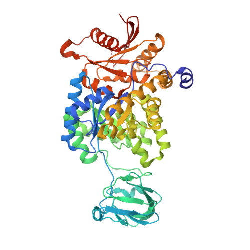
    Ehrlich's pioneering chemotherapeutic experiments published in 1904 (Ehrlich, P., and Shiga, K. (1904) Berlin Klin. Wochenschrift 20, 329-362) described the efficacy of a series of dye molecules including trypan blue and trypan red to eliminate trypanosome infections in mice. The molecular structures of the dyes provided a starting point for the synthesis of suramin, which was developed and used as a trypanocidal drug in 1916 and is still in clinical use. Despite the biological importance of these dye-like molecules, the mode of action on trypanosomes has remained elusive. Here we present crystal structures of suramin and three related dyes in complex with pyruvate kinases from Leishmania mexicana or from Trypanosoma cruzi. The phenyl sulfonate groups of all four molecules (suramin, Ponceau S, acid blue 80, and benzothiazole-2,5-disulfonic acid) bind in the position of ADP/ATP at the active sites of the pyruvate kinases (PYKs). The binding positions in the two different trypanosomatid PYKs are nearly identical. We show that suramin competitively inhibits PYKs from humans (muscle, tumor, and liver isoenzymes, K(i) = 1.1-17 μM), T. cruzi (K(i) = 108 μM), and L. mexicana (K(i) = 116 μM), all of which have similar active sites. Synergistic effects were observed when examining suramin inhibition in the presence of an allosteric effector molecule, whereby IC(50) values decreased up to 2-fold for both trypanosomatid and human PYKs. These kinetic and structural analyses provide insight into the promiscuous inhibition observed for suramin and into the mode of action of the dye-like molecules used in Ehrlich's original experiments.


  • Organizational Affiliation

    Structural Biochemistry Group, Institute of Structural and Molecular Biology, University of Edinburgh, King's Buildings, Mayfield Road, Edinburgh EH9 3JR, Scotland, United Kingdom.


Macromolecules
Find similar proteins by:  (by identity cutoff)  |  3D Structure
Entity ID: 1
MoleculeChains Sequence LengthOrganismDetailsImage
Pyruvate kinase
A, B
498Leishmania mexicanaMutation(s): 0 
Gene Names: PYK
EC: 2.7.1.40
UniProt
Find proteins for Q27686 (Leishmania mexicana)
Explore Q27686 
Go to UniProtKB:  Q27686
Entity Groups  
Sequence Clusters30% Identity50% Identity70% Identity90% Identity95% Identity100% Identity
UniProt GroupQ27686
Sequence Annotations
Expand
  • Reference Sequence
Small Molecules
Ligands 4 Unique
IDChains Name / Formula / InChI Key2D Diagram3D Interactions
SVR
Query on SVR

Download Ideal Coordinates CCD File 
G [auth B]8,8'-[CARBONYLBIS[IMINO-3,1-PHENYLENECARBONYLIMINO(4-METHYL-3,1-PHENYLENE)CARBONYLIMINO]]BIS-1,3,5-NAPHTHALENETRISULFON IC ACID
C51 H40 N6 O23 S6
FIAFUQMPZJWCLV-UHFFFAOYSA-N
PTK
Query on PTK

Download Ideal Coordinates CCD File 
C [auth A]pyrene-1,3,6,8-tetrasulfonic acid
C16 H10 O12 S4
CZLSHVQVNDDHDQ-UHFFFAOYSA-N
GOL
Query on GOL

Download Ideal Coordinates CCD File 
D [auth A],
J [auth B]
GLYCEROL
C3 H8 O3
PEDCQBHIVMGVHV-UHFFFAOYSA-N
K
Query on K

Download Ideal Coordinates CCD File 
E [auth A],
F [auth A],
H [auth B],
I [auth B]
POTASSIUM ION
K
NPYPAHLBTDXSSS-UHFFFAOYSA-N
Binding Affinity Annotations 
IDSourceBinding Affinity
SVR PDBBind:  3PP7 Ki: 1.16e+5 (nM) from 1 assay(s)
Experimental Data & Validation

Experimental Data

  • Method: X-RAY DIFFRACTION
  • Resolution: 2.35 Å
  • R-Value Free: 0.231 
  • R-Value Work: 0.191 
  • R-Value Observed: 0.193 
  • Space Group: I 2 2 2
Unit Cell:
Length ( Å )Angle ( ˚ )
a = 123.77α = 90
b = 129.48β = 90
c = 165.75γ = 90
Software Package:
Software NamePurpose
ADSCdata collection
REFMACrefinement
MOSFLMdata reduction
SCALAdata scaling

Structure Validation

View Full Validation Report


Ligand Structure Quality Assessment 


Entry History 

Deposition Data

Revision History  (Full details and data files)

  • Version 1.0: 2010-12-15
    Type: Initial release
  • Version 1.1: 2011-07-13
    Changes: Version format compliance
  • Version 1.2: 2011-08-03
    Changes: Database references
  • Version 1.3: 2011-09-21
    Changes: Database references
  • Version 1.4: 2023-09-06
    Changes: Data collection, Database references, Derived calculations, Refinement description, Structure summary