3POZ

EGFR Kinase domain complexed with tak-285


Experimental Data Snapshot

  • Method: X-RAY DIFFRACTION
  • Resolution: 1.50 Å
  • R-Value Free: 0.243 
  • R-Value Work: 0.219 
  • R-Value Observed: 0.220 

wwPDB Validation   3D Report Full Report


This is version 1.2 of the entry. See complete history


Literature

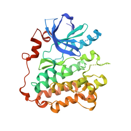
Structural Analysis of the Mechanism of Inhibition and Allosteric Activation of the Kinase Domain of HER2 Protein.

Aertgeerts, K.Skene, R.Yano, J.Sang, B.C.Zou, H.Snell, G.Jennings, A.Iwamoto, K.Habuka, N.Hirokawa, A.Ishikawa, T.Tanaka, T.Miki, H.Ohta, Y.Sogabe, S.

(2011) J Biol Chem 286: 18756-18765

  • DOI: https://doi.org/10.1074/jbc.M110.206193
  • Primary Citation of Related Structures:  
    3POZ, 3PP0

  • PubMed Abstract: 

    Aberrant signaling of ErbB family members human epidermal growth factor 2 (HER2) and epidermal growth factor receptor (EGFR) is implicated in many human cancers, and HER2 expression is predictive of human disease recurrence and prognosis. Small molecule kinase inhibitors of EGFR and of both HER2 and EGFR have received approval for the treatment of cancer. We present the first high resolution crystal structure of the kinase domain of HER2 in complex with a selective inhibitor to understand protein activation, inhibition, and function at the molecular level. HER2 kinase domain crystallizes as a dimer and suggests evidence for an allosteric mechanism of activation comparable with previously reported activation mechanisms for EGFR and HER4. A unique Gly-rich region in HER2 following the α-helix C is responsible for increased conformational flexibility within the active site and could explain the low intrinsic catalytic activity previously reported for HER2. In addition, we solved the crystal structure of the kinase domain of EGFR in complex with a HER2/EGFR dual inhibitor (TAK-285). Comparison with previously reported inactive and active EGFR kinase domain structures gave insight into the mechanism of HER2 and EGFR inhibition and may help guide the design and development of new cancer drugs with improved potency and selectivity.


  • Organizational Affiliation

    Takeda San Diego Inc, 10410 Science Center Drive, San Diego, California 92121, USA. kaertgeerts@takedasd.com


Macromolecules
Find similar proteins by:  (by identity cutoff)  |  3D Structure
Entity ID: 1
MoleculeChains Sequence LengthOrganismDetailsImage
Epidermal growth factor receptor327Homo sapiensMutation(s): 0 
Gene Names: EGFRERBB1
EC: 2.7.10.1
UniProt & NIH Common Fund Data Resources
Find proteins for P00533 (Homo sapiens)
Explore P00533 
Go to UniProtKB:  P00533
PHAROS:  P00533
GTEx:  ENSG00000146648 
Entity Groups  
Sequence Clusters30% Identity50% Identity70% Identity90% Identity95% Identity100% Identity
UniProt GroupP00533
Sequence Annotations
Expand
  • Reference Sequence
Small Molecules
Ligands 2 Unique
IDChains Name / Formula / InChI Key2D Diagram3D Interactions
03P
Query on 03P

Download Ideal Coordinates CCD File 
E [auth A]N-{2-[4-({3-chloro-4-[3-(trifluoromethyl)phenoxy]phenyl}amino)-5H-pyrrolo[3,2-d]pyrimidin-5-yl]ethyl}-3-hydroxy-3-methylbutanamide
C26 H25 Cl F3 N5 O3
ZYQXEVJIFYIBHZ-UHFFFAOYSA-N
SO4
Query on SO4

Download Ideal Coordinates CCD File 
B [auth A],
C [auth A],
D [auth A]
SULFATE ION
O4 S
QAOWNCQODCNURD-UHFFFAOYSA-L
Binding Affinity Annotations 
IDSourceBinding Affinity
03P BindingDB:  3POZ IC50: min: 1, max: 23 (nM) from 3 assay(s)
Binding MOAD:  3POZ IC50: 23 (nM) from 1 assay(s)
PDBBind:  3POZ IC50: 23 (nM) from 1 assay(s)
Experimental Data & Validation

Experimental Data

  • Method: X-RAY DIFFRACTION
  • Resolution: 1.50 Å
  • R-Value Free: 0.243 
  • R-Value Work: 0.219 
  • R-Value Observed: 0.220 
  • Space Group: P 21 21 21
Unit Cell:
Length ( Å )Angle ( ˚ )
a = 46.806α = 90
b = 68.881β = 90
c = 104.558γ = 90
Software Package:
Software NamePurpose
d*TREKdata scaling
d*TREKdata reduction
MOLREPphasing
REFMACrefinement
Cootmodel building

Structure Validation

View Full Validation Report


Entry History 

Deposition Data

Revision History  (Full details and data files)

  • Version 1.0: 2011-03-30
    Type: Initial release
  • Version 1.1: 2011-07-13
    Changes: Version format compliance
  • Version 1.2: 2023-09-06
    Changes: Data collection, Database references, Derived calculations, Refinement description