3PO7

Human monoamine oxidase B in complex with zonisamide


Experimental Data Snapshot

  • Method: X-RAY DIFFRACTION
  • Resolution: 1.80 Å
  • R-Value Free: 0.212 
  • R-Value Work: 0.182 
  • R-Value Observed: 0.183 

wwPDB Validation   3D Report Full Report


Ligand Structure Quality Assessment 


This is version 1.3 of the entry. See complete history


Literature

Interactions of monoamine oxidases with the antiepileptic drug zonisamide: specificity of inhibition and structure of the human monoamine oxidase B complex

Binda, C.Aldeco, M.Mattevi, A.Edmondson, D.E.

(2011) J Med Chem 54: 909-912

  • DOI: https://doi.org/10.1021/jm101359c
  • Primary Citation of Related Structures:  
    3PO7

  • PubMed Abstract: 

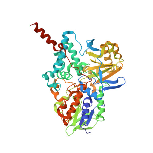
    The binding of zonisamide to purified, recombinant monoamine oxidases (MAOs) has been investigated. It is a competitive inhibitor of human MAO B (K(i) = 3.1 ± 0.3 μM), of rat MAO B (K(i) = 2.9 ± 0.5 μM), and of zebrafish MAO (K(i) = 30.8 ± 5.3 μM). No inhibition is observed with purified human or rat MAO A. The 1.8 Å structure of the MAO B complex demonstrates that it binds within the substrate cavity.


  • Organizational Affiliation

    Department Genetics and Microbiology, University of Pavia, via Ferrata 1, Pavia 27100, Italy.


Macromolecules
Find similar proteins by:  (by identity cutoff)  |  3D Structure
Entity ID: 1
MoleculeChains Sequence LengthOrganismDetailsImage
Amine oxidase [flavin-containing] B
A, B
520Homo sapiensMutation(s): 0 
Gene Names: MAOB
EC: 1.4.3.4
Membrane Entity: Yes 
UniProt & NIH Common Fund Data Resources
Find proteins for P27338 (Homo sapiens)
Explore P27338 
Go to UniProtKB:  P27338
PHAROS:  P27338
GTEx:  ENSG00000069535 
Entity Groups  
Sequence Clusters30% Identity50% Identity70% Identity90% Identity95% Identity100% Identity
UniProt GroupP27338
Sequence Annotations
Expand
  • Reference Sequence
Small Molecules
Binding Affinity Annotations 
IDSourceBinding Affinity
ZON BindingDB:  3PO7 Ki: min: 2900, max: 3100 (nM) from 2 assay(s)
Binding MOAD:  3PO7 Ki: 3100 (nM) from 1 assay(s)
Experimental Data & Validation

Experimental Data

  • Method: X-RAY DIFFRACTION
  • Resolution: 1.80 Å
  • R-Value Free: 0.212 
  • R-Value Work: 0.182 
  • R-Value Observed: 0.183 
  • Space Group: C 2 2 2
Unit Cell:
Length ( Å )Angle ( ˚ )
a = 131.4α = 90
b = 222.6β = 90
c = 86.19γ = 90
Software Package:
Software NamePurpose
DNAdata collection
AMoREphasing
REFMACrefinement
MOSFLMdata reduction
SCALAdata scaling

Structure Validation

View Full Validation Report


Ligand Structure Quality Assessment 


Entry History 

Deposition Data

Revision History  (Full details and data files)

  • Version 1.0: 2011-01-12
    Type: Initial release
  • Version 1.1: 2011-07-13
    Changes: Version format compliance
  • Version 1.2: 2014-03-05
    Changes: Database references
  • Version 1.3: 2023-11-01
    Changes: Data collection, Database references, Derived calculations, Refinement description