3PNB

Phosphopantetheine Adenylyltransferase from Mycobacterium tuberculosis in complex with coenzyme A


Experimental Data Snapshot

  • Method: X-RAY DIFFRACTION
  • Resolution: 2.11 Å
  • R-Value Free: 0.248 
  • R-Value Work: 0.215 
  • R-Value Observed: 0.216 

wwPDB Validation   3D Report Full Report


Ligand Structure Quality Assessment 


This is version 1.2 of the entry. See complete history


Literature

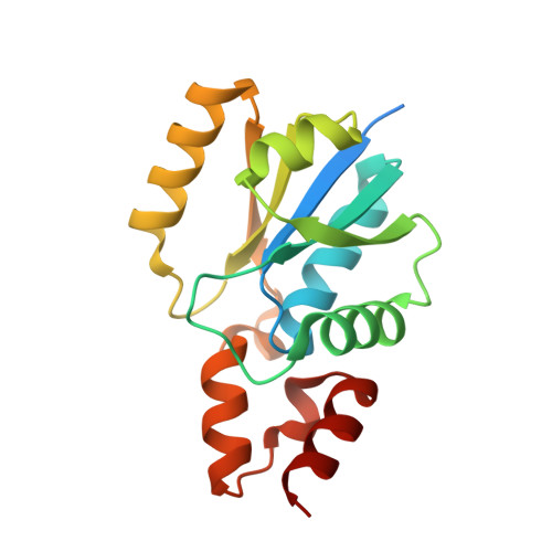
Structure of Mycobacterium tuberculosisphosphopantetheine adenylyltransferase in complex with the feedback inhibitor CoA reveals only one active-site conformation.

Wubben, T.Mesecar, A.D.

(2011) Acta Crystallogr Sect F Struct Biol Cryst Commun 67: 541-545

  • DOI: https://doi.org/10.1107/S1744309111010761
  • Primary Citation of Related Structures:  
    3PNB

  • PubMed Abstract: 

    Phosphopantetheine adenylyltransferase (PPAT) catalyzes the penultimate step in the coenzyme A (CoA) biosynthetic pathway, reversibly transferring an adenylyl group from ATP to 4'-phosphopantetheine to form dephosphocoenzyme A (dPCoA). To complement recent biochemical and structural studies on Mycobacterium tuberculosis PPAT (MtPPAT) and to provide further insight into the feedback regulation of MtPPAT by CoA, the X-ray crystal structure of the MtPPAT enzyme in complex with CoA was determined to 2.11 Å resolution. Unlike previous X-ray crystal structures of PPAT-CoA complexes from other bacteria, which showed two distinct CoA conformations bound to the active site, only one conformation of CoA is observed in the MtPPAT-CoA complex.


  • Organizational Affiliation

    Department of Biochemistry and Molecular Genetics, University of Illinois at Chicago, Chicago, Illinois, USA.


Macromolecules
Find similar proteins by:  (by identity cutoff)  |  3D Structure
Entity ID: 1
MoleculeChains Sequence LengthOrganismDetailsImage
Phosphopantetheine adenylyltransferase
A, B, C, D
177Mycobacterium tuberculosisMutation(s): 0 
Gene Names: coaDkdtBRv2965cMT3043MTCY349.22u0002e
EC: 2.7.7.3
UniProt
Find proteins for P9WPA5 (Mycobacterium tuberculosis (strain ATCC 25618 / H37Rv))
Explore P9WPA5 
Go to UniProtKB:  P9WPA5
Entity Groups  
Sequence Clusters30% Identity50% Identity70% Identity90% Identity95% Identity100% Identity
UniProt GroupP9WPA5
Sequence Annotations
Expand
  • Reference Sequence
Experimental Data & Validation

Experimental Data

  • Method: X-RAY DIFFRACTION
  • Resolution: 2.11 Å
  • R-Value Free: 0.248 
  • R-Value Work: 0.215 
  • R-Value Observed: 0.216 
  • Space Group: P 3 2 1
Unit Cell:
Length ( Å )Angle ( ˚ )
a = 114.459α = 90
b = 114.459β = 90
c = 134.47γ = 120
Software Package:
Software NamePurpose
MD2data collection
PHASERphasing
REFMACrefinement
HKL-2000data reduction
HKL-2000data scaling

Structure Validation

View Full Validation Report


Ligand Structure Quality Assessment 


Entry History 

Deposition Data

Revision History  (Full details and data files)

  • Version 1.0: 2011-06-22
    Type: Initial release
  • Version 1.1: 2011-07-13
    Changes: Version format compliance
  • Version 1.2: 2024-02-21
    Changes: Data collection, Database references, Derived calculations