3PN7

Visualizing new hinges and a potential major source of compliance in the lever arm of myosin


Experimental Data Snapshot

  • Method: X-RAY DIFFRACTION
  • Resolution: 2.25 Å
  • R-Value Free: 0.241 
  • R-Value Work: 0.191 
  • R-Value Observed: 0.191 

wwPDB Validation   3D Report Full Report


This is version 1.2 of the entry. See complete history


Literature

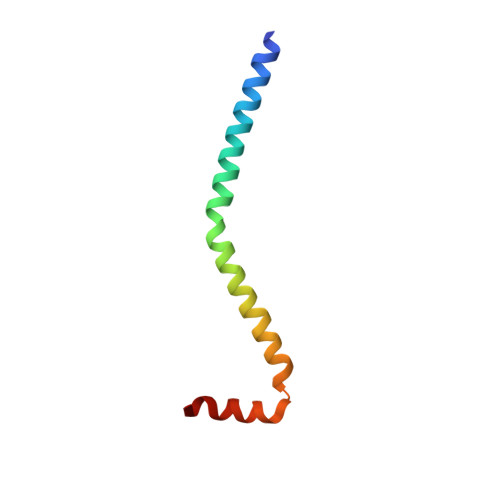
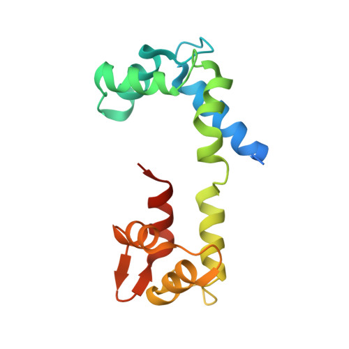
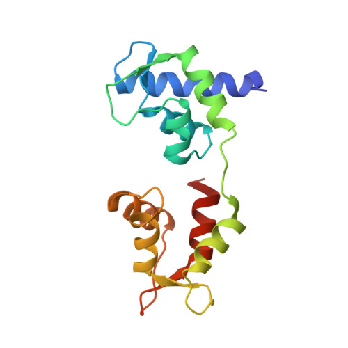
Visualizing key hinges and a potential major source of compliance in the lever arm of myosin.

Brown, J.H.Senthil Kumar, V.S.O'Neall-Hennessey, E.Reshetnikova, L.Robinson, H.Nguyen-McCarty, M.Szent-Gyorgyi, A.G.Cohen, C.

(2011) Proc Natl Acad Sci U S A 108: 114-119

  • DOI: https://doi.org/10.1073/pnas.1016288107
  • Primary Citation of Related Structures:  
    3PN7

  • PubMed Abstract: 

    We have determined the 2.3-Å-resolution crystal structure of a myosin light chain domain, corresponding to one type found in sea scallop catch ("smooth") muscle. This structure reveals hinges that may function in the "on" and "off" states of myosin. The molecule adopts two different conformations about the heavy chain "hook" and regulatory light chain (RLC) helix D. This conformational change results in extended and compressed forms of the lever arm whose lengths differ by 10 Å. The heavy chain hook and RLC helix D hinges could thus serve as a potential major and localized source of cross-bridge compliance during the contractile cycle. In addition, in one of the molecules of the crystal, part of the RLC N-terminal extension is seen in atomic detail and forms a one-turn alpha-helix that interacts with RLC helix D. This extension, whose sequence is highly variable in different myosins, may thus modulate the flexibility of the lever arm. Moreover, the relative proximity of the phosphorylation site to the helix D hinge suggests a potential role for conformational changes about this hinge in the transition between the on and off states of regulated myosins.


  • Organizational Affiliation

    Rosenstiel Basic Medical Sciences Research Center, Brandeis University, Waltham, MA 02454-9110, USA.


Macromolecules
Find similar proteins by:  (by identity cutoff)  |  3D Structure
Entity ID: 1
MoleculeChains Sequence LengthOrganismDetailsImage
Myosin heavy chain
A, D
69Placopecten magellanicusMutation(s): 0 
UniProt
Find proteins for Q26080 (Placopecten magellanicus)
Explore Q26080 
Go to UniProtKB:  Q26080
Entity Groups  
Sequence Clusters30% Identity50% Identity70% Identity90% Identity95% Identity100% Identity
UniProt GroupQ26080
Sequence Annotations
Expand
  • Reference Sequence
Find similar proteins by:  (by identity cutoff)  |  3D Structure
Entity ID: 2
MoleculeChains Sequence LengthOrganismDetailsImage
Myosin regulatory light chain
B, E
161Placopecten magellanicusMutation(s): 0 
UniProt
Find proteins for Q26069 (Placopecten magellanicus)
Explore Q26069 
Go to UniProtKB:  Q26069
Entity Groups  
Sequence Clusters30% Identity50% Identity70% Identity90% Identity95% Identity100% Identity
UniProt GroupQ26069
Sequence Annotations
Expand
  • Reference Sequence
Find similar proteins by:  (by identity cutoff)  |  3D Structure
Entity ID: 3
MoleculeChains Sequence LengthOrganismDetailsImage
Myosin essential light chain
C, F
156Placopecten magellanicusMutation(s): 0 
UniProt
Find proteins for Q26066 (Placopecten magellanicus)
Explore Q26066 
Go to UniProtKB:  Q26066
Entity Groups  
Sequence Clusters30% Identity50% Identity70% Identity90% Identity95% Identity100% Identity
UniProt GroupQ26066
Sequence Annotations
Expand
  • Reference Sequence
Experimental Data & Validation

Experimental Data

  • Method: X-RAY DIFFRACTION
  • Resolution: 2.25 Å
  • R-Value Free: 0.241 
  • R-Value Work: 0.191 
  • R-Value Observed: 0.191 
  • Space Group: P 1
Unit Cell:
Length ( Å )Angle ( ˚ )
a = 50.737α = 77.41
b = 68.779β = 85.94
c = 79.305γ = 73.61
Software Package:
Software NamePurpose
HKL-2000data collection
PHASERphasing
PHENIXrefinement
HKL-2000data reduction
HKL-2000data scaling

Structure Validation

View Full Validation Report


Entry History 

Revision History  (Full details and data files)

  • Version 1.0: 2011-01-05
    Type: Initial release
  • Version 1.1: 2011-07-13
    Changes: Version format compliance
  • Version 1.2: 2023-09-06
    Changes: Data collection, Database references, Derived calculations, Refinement description