3PN4

Crystal structure of Arabidopsis thaliana petide deformylase 1B (AtPDF1B) in complex with actinonin (crystallized in PEG-550-MME)


Experimental Data Snapshot

  • Method: X-RAY DIFFRACTION
  • Resolution: 1.90 Å
  • R-Value Free: 0.210 
  • R-Value Work: 0.182 
  • R-Value Observed: 0.183 

wwPDB Validation   3D Report Full Report


Ligand Structure Quality Assessment 


This is version 1.2 of the entry. See complete history


Literature

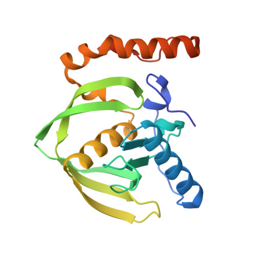
Trapping conformational States along ligand-binding dynamics of Peptide deformylase: the impact of induced fit on enzyme catalysis.

Fieulaine, S.Boularot, A.Artaud, I.Desmadril, M.Dardel, F.Meinnel, T.Giglione, C.

(2011) PLoS Biol 9: e1001066-e1001066

  • DOI: https://doi.org/10.1371/journal.pbio.1001066
  • Primary Citation of Related Structures:  
    3M6O, 3M6P, 3M6Q, 3M6R, 3O3J, 3PN2, 3PN3, 3PN4, 3PN5, 3PN6

  • PubMed Abstract: 

    For several decades, molecular recognition has been considered one of the most fundamental processes in biochemistry. For enzymes, substrate binding is often coupled to conformational changes that alter the local environment of the active site to align the reactive groups for efficient catalysis and to reach the transition state. Adaptive substrate recognition is a well-known concept; however, it has been poorly characterized at a structural level because of its dynamic nature. Here, we provide a detailed mechanism for an induced-fit process at atomic resolution. We take advantage of a slow, tight binding inhibitor-enzyme system, actinonin-peptide deformylase. Crystal structures of the initial open state and final closed state were solved, as well as those of several intermediate mimics captured during the process. Ligand-induced reshaping of a hydrophobic pocket drives closure of the active site, which is finally "zipped up" by additional binding interactions. Together with biochemical analyses, these data allow a coherent reconstruction of the sequence of events leading from the encounter complex to the key-lock binding state of the enzyme. A "movie" that reconstructs this entire process can be further extrapolated to catalysis.


  • Organizational Affiliation

    CNRS, ISV, UPR2355, Gif-sur-Yvette, France.


Macromolecules
Find similar proteins by:  (by identity cutoff)  |  3D Structure
Entity ID: 1
MoleculeChains Sequence LengthOrganismDetailsImage
Peptide deformylase 1B, chloroplastic193Arabidopsis thalianaMutation(s): 0 
Gene Names: DEF2PDF1B
EC: 3.5.1.88
UniProt
Find proteins for Q9FUZ2 (Arabidopsis thaliana)
Explore Q9FUZ2 
Go to UniProtKB:  Q9FUZ2
Entity Groups  
Sequence Clusters30% Identity50% Identity70% Identity90% Identity95% Identity100% Identity
UniProt GroupQ9FUZ2
Sequence Annotations
Expand
  • Reference Sequence
Experimental Data & Validation

Experimental Data

  • Method: X-RAY DIFFRACTION
  • Resolution: 1.90 Å
  • R-Value Free: 0.210 
  • R-Value Work: 0.182 
  • R-Value Observed: 0.183 
  • Space Group: P 43 21 2
Unit Cell:
Length ( Å )Angle ( ˚ )
a = 56.57α = 90
b = 56.57β = 90
c = 146.11γ = 90
Software Package:
Software NamePurpose
ADSCdata collection
REFMACrefinement
XDSdata reduction
XDSdata scaling
REFMACphasing

Structure Validation

View Full Validation Report


Ligand Structure Quality Assessment 


Entry History 

Deposition Data

Revision History  (Full details and data files)

  • Version 1.0: 2011-06-08
    Type: Initial release
  • Version 1.1: 2011-07-13
    Changes: Version format compliance
  • Version 1.2: 2023-09-06
    Changes: Data collection, Database references, Derived calculations, Refinement description