3PHS

Crystal Structure of GBS52, the minor pilin in gram-positive pathogen Streptococcus agalactiae


Experimental Data Snapshot

  • Method: X-RAY DIFFRACTION
  • Resolution: 1.80 Å
  • R-Value Free: 0.230 
  • R-Value Work: 0.199 
  • R-Value Observed: 0.201 

wwPDB Validation   3D Report Full Report


This is version 1.1 of the entry. See complete history


Literature

An IgG-like domain in the minor pilin GBS52 of Streptococcus agalactiae mediates lung epithelial cell adhesion.

Krishnan, V.Gaspar, A.H.Ye, N.Mandlik, A.Ton-That, H.Narayana, S.V.

(2007) Structure 15: 893-903

  • DOI: https://doi.org/10.1016/j.str.2007.06.015
  • Primary Citation of Related Structures:  
    3PHS

  • PubMed Abstract: 

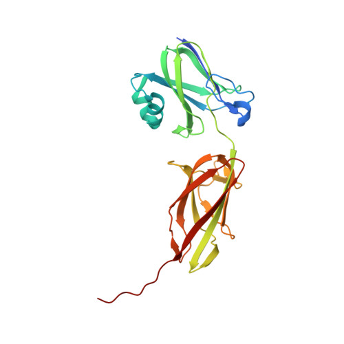
    Streptococcus agalactiae is the leading cause of neonatal pneumonia, sepsis, and meningitis. The pathogen assembles heterotrimeric pilus structures on its surface; however, their function in pathogenesis is poorly understood. We report here the crystal structure of the pilin GBS52, which reveals two IgG-like fold domains, N1 and N2. Each domain is comprised of seven antiparallel beta strands, an arrangement similar to the fold observed in the Staphylococcus aureus adhesin Cna. Consistent with its role as an adhesin, deletion of gbs52 gene significantly reduces bacterial adherence to pulmonary epithelial cells. Moreover, latex beads linked to the GBS52 protein adhere to pulmonary but not to many other epithelial cells; binding to the former is specifically inhibited by antibodies against GBS52. Nonetheless, substantial binding is only observed with N2 domain-conjugated beads. This study presents the structure of a Gram-positive pilin that utilizes a distinct IgG fold variant to mediate pathogen adherence to a specific tissue.


  • Organizational Affiliation

    School of Optometry and Center for Biophysical Sciences and Engineering, University of Alabama at Birmingham, AL 35294, USA.


Macromolecules
Find similar proteins by:  (by identity cutoff)  |  3D Structure
Entity ID: 1
MoleculeChains Sequence LengthOrganismDetailsImage
Cell wall surface anchor family protein249Streptococcus agalactiae 2603V/RMutation(s): 0 
Gene Names: GBS052SAG0646
UniProt
Find proteins for Q8E0S8 (Streptococcus agalactiae serotype V (strain ATCC BAA-611 / 2603 V/R))
Explore Q8E0S8 
Go to UniProtKB:  Q8E0S8
Entity Groups  
Sequence Clusters30% Identity50% Identity70% Identity90% Identity95% Identity100% Identity
UniProt GroupQ8E0S8
Sequence Annotations
Expand
  • Reference Sequence
Experimental Data & Validation

Experimental Data

  • Method: X-RAY DIFFRACTION
  • Resolution: 1.80 Å
  • R-Value Free: 0.230 
  • R-Value Work: 0.199 
  • R-Value Observed: 0.201 
  • Space Group: P 1 21 1
Unit Cell:
Length ( Å )Angle ( ˚ )
a = 41.481α = 90
b = 52.222β = 101.79
c = 61.006γ = 90
Software Package:
Software NamePurpose
SOLVEphasing
REFMACrefinement
d*TREKdata reduction
d*TREKdata scaling

Structure Validation

View Full Validation Report


Entry History 

Deposition Data

Revision History  (Full details and data files)

  • Version 1.0: 2010-11-17
    Type: Initial release
  • Version 1.1: 2011-07-13
    Changes: Version format compliance