3PFP

Structure of 3-deoxy-D-arabino-heptulosonate 7-phosphate synthase from Mycobacterium tuberculosis in complex with an active site inhibitor


Experimental Data Snapshot

  • Method: X-RAY DIFFRACTION
  • Resolution: 2.35 Å
  • R-Value Free: 0.196 
  • R-Value Work: 0.162 
  • R-Value Observed: 0.164 

wwPDB Validation   3D Report Full Report


Ligand Structure Quality Assessment 


This is version 1.3 of the entry. See complete history


Literature

Potent inhibitors of a shikimate pathway enzyme from Mycobacterium tuberculosis: combining mechanism- and modeling-based design

Reichau, S.Jiao, W.Walker, S.R.Hutton, R.D.Baker, E.N.Parker, E.J.

(2011) J Biol Chem 286: 16197-16207

  • DOI: https://doi.org/10.1074/jbc.M110.211649
  • Primary Citation of Related Structures:  
    3PFP

  • PubMed Abstract: 

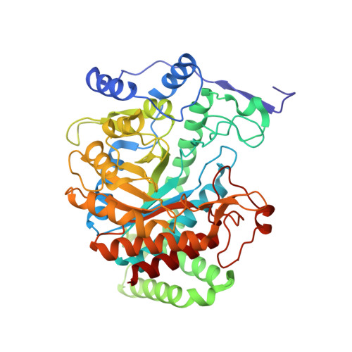
    Tuberculosis remains a serious global health threat, with the emergence of multidrug-resistant strains highlighting the urgent need for novel antituberculosis drugs. The enzyme 3-deoxy-D-arabino-heptulosonate 7-phosphate synthase (DAH7PS) catalyzes the first step of the shikimate pathway for the biosynthesis of aromatic compounds. This pathway has been shown to be essential in Mycobacterium tuberculosis, the pathogen responsible for tuberculosis. DAH7PS catalyzes a condensation reaction between P-enolpyruvate and erythrose 4-phosphate to give 3-deoxy-D-arabino-heptulosonate 7-phosphate. The enzyme reaction mechanism is proposed to include a tetrahedral intermediate, which is formed by attack of an active site water on the central carbon of P-enolpyruvate during the course of the reaction. Molecular modeling of this intermediate into the active site reported in this study shows a configurational preference consistent with water attack from the re face of P-enolpyruvate. Based on this model, we designed and synthesized an inhibitor of DAH7PS that mimics this reaction intermediate. Both enantiomers of this intermediate mimic were potent inhibitors of M. tuberculosis DAH7PS, with inhibitory constants in the nanomolar range. The crystal structure of the DAH7PS-inhibitor complex was solved to 2.35 Å. Both the position of the inhibitor and the conformational changes of active site residues observed in this structure correspond closely to the predictions from the intermediate modeling. This structure also identifies a water molecule that is located in the appropriate position to attack the re face of P-enolpyruvate during the course of the reaction, allowing the catalytic mechanism for this enzyme to be clearly defined.


  • Organizational Affiliation

    Biomolecular Interaction Centre and Department of Chemistry, University of Canterbury, Christchurch, New Zealand.


Macromolecules
Find similar proteins by:  (by identity cutoff)  |  3D Structure
Entity ID: 1
MoleculeChains Sequence LengthOrganismDetailsImage
Probable 3-deoxy-D-arabino-heptulosonate 7-phosphate synthase AroG
A, B
464Mycobacterium tuberculosisMutation(s): 0 
Gene Names: aroGRv2178c
EC: 2.5.1.54
UniProt
Find proteins for O53512 (Mycobacterium tuberculosis (strain ATCC 25618 / H37Rv))
Explore O53512 
Go to UniProtKB:  O53512
Entity Groups  
Sequence Clusters30% Identity50% Identity70% Identity90% Identity95% Identity100% Identity
UniProt GroupO53512
Sequence Annotations
Expand
  • Reference Sequence
Small Molecules
Ligands 5 Unique
IDChains Name / Formula / InChI Key2D Diagram3D Interactions
036
Query on 036

Download Ideal Coordinates CCD File 
E [auth A],
G [auth B]
(2R)-2,7-bis(phosphonooxy)heptanoic acid
C7 H16 O10 P2
OZYHUBGACYJJJF-ZCFIWIBFSA-N
035
Query on 035

Download Ideal Coordinates CCD File 
D [auth A],
J [auth B]
(2S)-2,7-bis(phosphonooxy)heptanoic acid
C7 H16 O10 P2
OZYHUBGACYJJJF-LURJTMIESA-N
SO4
Query on SO4

Download Ideal Coordinates CCD File 
I [auth B]SULFATE ION
O4 S
QAOWNCQODCNURD-UHFFFAOYSA-L
MN
Query on MN

Download Ideal Coordinates CCD File 
C [auth A],
F [auth B]
MANGANESE (II) ION
Mn
WAEMQWOKJMHJLA-UHFFFAOYSA-N
CL
Query on CL

Download Ideal Coordinates CCD File 
H [auth B]CHLORIDE ION
Cl
VEXZGXHMUGYJMC-UHFFFAOYSA-M
Experimental Data & Validation

Experimental Data

  • Method: X-RAY DIFFRACTION
  • Resolution: 2.35 Å
  • R-Value Free: 0.196 
  • R-Value Work: 0.162 
  • R-Value Observed: 0.164 
  • Space Group: P 32 2 1
Unit Cell:
Length ( Å )Angle ( ˚ )
a = 203.579α = 90
b = 203.579β = 90
c = 66.49γ = 120
Software Package:
Software NamePurpose
SCALAdata scaling
REFMACrefinement
PDB_EXTRACTdata extraction
ADSCdata collection
MOSFLMdata reduction

Structure Validation

View Full Validation Report


Ligand Structure Quality Assessment 


Entry History 

Deposition Data

Revision History  (Full details and data files)

  • Version 1.0: 2011-03-16
    Type: Initial release
  • Version 1.1: 2011-07-13
    Changes: Version format compliance
  • Version 1.2: 2012-02-22
    Changes: Database references, Structure summary
  • Version 1.3: 2023-11-01
    Changes: Data collection, Database references, Derived calculations, Refinement description