3PDN

Crystal structure of SmyD3 in complex with methyltransferase inhibitor sinefungin


Experimental Data Snapshot

  • Method: X-RAY DIFFRACTION
  • Resolution: 1.70 Å
  • R-Value Free: 0.189 
  • R-Value Work: 0.155 
  • R-Value Observed: 0.157 

wwPDB Validation   3D Report Full Report


Ligand Structure Quality Assessment 


This is version 1.2 of the entry. See complete history


Literature

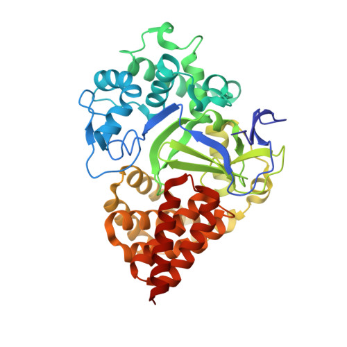
Structural Insights into the Autoinhibition and Posttranslational Activation of Histone Methyltransferase SmyD3.

Sirinupong, N.Brunzelle, J.Doko, E.Yang, Z.

(2011) J Mol Biol 406: 149-159

  • DOI: https://doi.org/10.1016/j.jmb.2010.12.014
  • Primary Citation of Related Structures:  
    3PDN

  • PubMed Abstract: 

    The SmyD family represents a new class of chromatin regulators that is important in heart and skeletal muscle development. However, the critical questions regarding how they are regulated posttranslationally remain largely unknown. We previously suggested that the histone methyltransferase activity of SmyD1, a vital myogenic regulator, appears to be regulated by autoinhibition and that the possible hinge motion of the conserved C-terminal domain (CTD) might be central to the maintenance and release of the autoinhibition. However, the lack of direct evidence of the hinge motion has limited our further understanding of this autoinhibitory mechanism. Here, we report the crystal structure of full-length SmyD3 in complex with the methyltransferase inhibitor sinefungin at 1.7 Å. SmyD3 has a two-lobed structure with the substrate binding cleft located at the bottom of a  15-Å-deep crevice formed between the N- and C-terminal lobes. Comparison of SmyD3 and SmyD1 clearly suggests that the CTD can undergo a large hinge-bending motion that defines two distinct conformations: SmyD3 adopts a closed conformation with the CTD partially blocking the substrate binding cleft; in contrast, SmyD1 appears to represent an open form, where the CTD swings out by ∼12 Å from the N-terminal lobe, forming an open cleft with the active site completely exposed. Overall, these findings provide novel structural insights into the mechanism that modulates the activity of the SmyD proteins and support the observation that a posttranslational activation, such as by molecular chaperon Hsp90, is required to potentiate the proteins.


  • Organizational Affiliation

    Department of Biochemistry and Molecular Biology, Wayne State University School of Medicine, Detroit, MI 48201, USA.


Macromolecules
Find similar proteins by:  (by identity cutoff)  |  3D Structure
Entity ID: 1
MoleculeChains Sequence LengthOrganismDetailsImage
SET and MYND domain-containing protein 3428Homo sapiensMutation(s): 0 
Gene Names: SMYD3ZMYND1ZNFN3A1
EC: 2.1.1.43
UniProt & NIH Common Fund Data Resources
Find proteins for Q9H7B4 (Homo sapiens)
Explore Q9H7B4 
Go to UniProtKB:  Q9H7B4
PHAROS:  Q9H7B4
GTEx:  ENSG00000185420 
Entity Groups  
Sequence Clusters30% Identity50% Identity70% Identity90% Identity95% Identity100% Identity
UniProt GroupQ9H7B4
Sequence Annotations
Expand
  • Reference Sequence
Small Molecules
Ligands 4 Unique
IDChains Name / Formula / InChI Key2D Diagram3D Interactions
SFG
Query on SFG

Download Ideal Coordinates CCD File 
B [auth A]SINEFUNGIN
C15 H23 N7 O5
LMXOHSDXUQEUSF-YECHIGJVSA-N
GOL
Query on GOL

Download Ideal Coordinates CCD File 
C [auth A]
D [auth A]
E [auth A]
F [auth A]
G [auth A]
C [auth A],
D [auth A],
E [auth A],
F [auth A],
G [auth A],
H [auth A]
GLYCEROL
C3 H8 O3
PEDCQBHIVMGVHV-UHFFFAOYSA-N
ZN
Query on ZN

Download Ideal Coordinates CCD File 
I [auth A],
J [auth A],
K [auth A]
ZINC ION
Zn
PTFCDOFLOPIGGS-UHFFFAOYSA-N
MG
Query on MG

Download Ideal Coordinates CCD File 
L [auth A],
M [auth A]
MAGNESIUM ION
Mg
JLVVSXFLKOJNIY-UHFFFAOYSA-N
Experimental Data & Validation

Experimental Data

  • Method: X-RAY DIFFRACTION
  • Resolution: 1.70 Å
  • R-Value Free: 0.189 
  • R-Value Work: 0.155 
  • R-Value Observed: 0.157 
  • Space Group: P 21 21 21
Unit Cell:
Length ( Å )Angle ( ˚ )
a = 61.24α = 90
b = 66.246β = 90
c = 107.384γ = 90
Software Package:
Software NamePurpose
HKL-2000data collection
SOLVEphasing
CNSrefinement
HKL-2000data reduction
SCALEPACKdata scaling
BUSTERrefinement

Structure Validation

View Full Validation Report


Ligand Structure Quality Assessment 


Entry History 

Deposition Data

Revision History  (Full details and data files)

  • Version 1.0: 2010-11-17
    Type: Initial release
  • Version 1.1: 2011-07-13
    Changes: Version format compliance
  • Version 1.2: 2024-02-21
    Changes: Data collection, Database references, Derived calculations