3PAB

Crystal Structure of H2-Kb in complex with a mutant of the chicken ovalbumin epitope OVA-E1


Experimental Data Snapshot

  • Method: X-RAY DIFFRACTION
  • Resolution: 2.20 Å
  • R-Value Free: 0.260 
  • R-Value Work: 0.193 
  • R-Value Observed: 0.196 

wwPDB Validation   3D Report Full Report


This is version 1.3 of the entry. See complete history


Literature

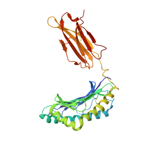
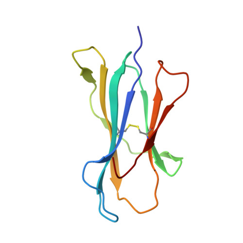
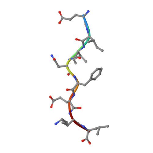
Affinity thresholds for naive CD8+ CTL activation by peptides and engineered influenza A viruses

Denton, A.E.Wesselingh, R.Gras, S.Guillonneau, C.Olson, M.R.Mintern, J.D.Zeng, W.Jackson, D.C.Rossjohn, J.Hodgkin, P.D.Doherty, P.C.Turner, S.J.

(2011) J Immunol 187: 5733-5744

  • DOI: https://doi.org/10.4049/jimmunol.1003937
  • Primary Citation of Related Structures:  
    3P9L, 3P9M, 3PAB

  • PubMed Abstract: 

    High-avidity interactions between TCRs and peptide + class I MHC (pMHCI) epitopes drive CTL activation and expansion. Intriguing questions remain concerning the constraints determining optimal TCR/pMHCI binding. The present analysis uses the TCR transgenic OT-I model to assess how varying profiles of TCR/pMHCI avidity influence naive CTL proliferation and the acquisition of effector function following exposure to the cognate H-2K(b)/OVA(257-264) (SIINFEKL) epitope and to mutants provided as peptide or in engineered influenza A viruses. Stimulating naive OT-I CD8(+) T cells in vitro with SIINFEKL induced full CTL proliferation and differentiation that was largely independent of any need for costimulation. By contrast, in vitro activation with the low-affinity EIINFEKL or SIIGFEKL ligands depended on the provision of IL-2 and other costimulatory signals. Importantly, although they did generate potent endogenous responses, infection of mice with influenza A viruses expressing these same OVA(257) variants failed to induce the activation of adoptively transferred naive OT-I CTLps, an effect that was only partially overcome by priming with a lipopeptide vaccine. Subsequent structural and biophysical analysis of H2-K(b)OVA(257), H2-K(b)E1, and H2-K(b)G4 established that these variations introduce small changes at the pMHCI interface and decrease epitope stability in ways that would likely impact cell surface presentation and recognition. Overall, it seems that there is an activation threshold for naive CTLps, that minimal alterations in peptide sequence can have profound effects, and that the antigenic requirements for the in vitro and in vivo induction of CTL proliferation and effector function differ substantially.


  • Organizational Affiliation

    Department of Microbiology and Immunology, University of Melbourne, Parkville, Victoria 3010, Australia.


Macromolecules
Find similar proteins by:  (by identity cutoff)  |  3D Structure
Entity ID: 1
MoleculeChains Sequence LengthOrganismDetailsImage
H-2 class I histocompatibility antigen, K-B alpha chain
A, D
279Mus musculusMutation(s): 0 
Gene Names: H2-K1H2-K
UniProt
Find proteins for P01901 (Mus musculus)
Explore P01901 
Go to UniProtKB:  P01901
Entity Groups  
Sequence Clusters30% Identity50% Identity70% Identity90% Identity95% Identity100% Identity
UniProt GroupP01901
Sequence Annotations
Expand
  • Reference Sequence
Find similar proteins by:  (by identity cutoff)  |  3D Structure
Entity ID: 2
MoleculeChains Sequence LengthOrganismDetailsImage
Beta-2-microglobulin
B, E
99Mus musculusMutation(s): 0 
Gene Names: B2m
UniProt & NIH Common Fund Data Resources
Find proteins for P01887 (Mus musculus)
Explore P01887 
Go to UniProtKB:  P01887
IMPC:  MGI:88127
Entity Groups  
Sequence Clusters30% Identity50% Identity70% Identity90% Identity95% Identity100% Identity
UniProt GroupP01887
Sequence Annotations
Expand
  • Reference Sequence

Find similar proteins by:  Sequence   |   3D Structure  

Entity ID: 3
MoleculeChains Sequence LengthOrganismDetailsImage
Ovalbumin epitope, EIINFEKL
C, F
8N/AMutation(s): 1 
UniProt
Find proteins for P01012 (Gallus gallus)
Explore P01012 
Go to UniProtKB:  P01012
Entity Groups  
UniProt GroupP01012
Sequence Annotations
Expand
  • Reference Sequence
Experimental Data & Validation

Experimental Data

  • Method: X-RAY DIFFRACTION
  • Resolution: 2.20 Å
  • R-Value Free: 0.260 
  • R-Value Work: 0.193 
  • R-Value Observed: 0.196 
  • Space Group: P 1 21 1
Unit Cell:
Length ( Å )Angle ( ˚ )
a = 66.683α = 90
b = 88.818β = 111.38
c = 89.121γ = 90
Software Package:
Software NamePurpose
XSCALEdata scaling
PHASERphasing
REFMACrefinement
PDB_EXTRACTdata extraction
PHENIXrefinement

Structure Validation

View Full Validation Report


Entry History 

Deposition Data

Revision History  (Full details and data files)

  • Version 1.0: 2011-10-19
    Type: Initial release
  • Version 1.1: 2011-11-09
    Changes: Database references
  • Version 1.2: 2013-06-19
    Changes: Database references
  • Version 1.3: 2023-11-01
    Changes: Data collection, Database references, Refinement description