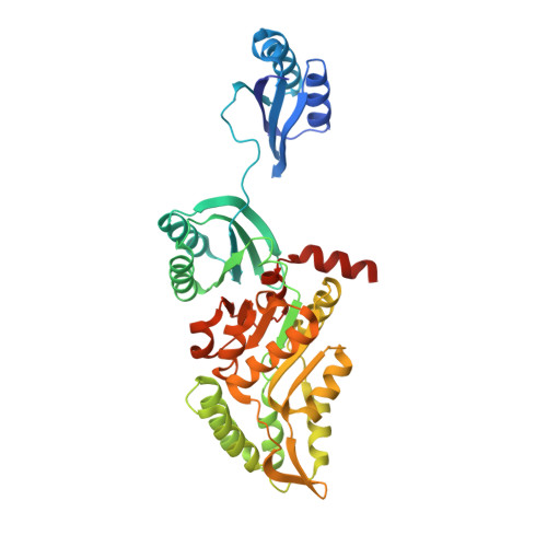
SAD phasing using iodide ions in a high-throughput structural genomics environment.
Abendroth, J., Gardberg, A.S., Robinson, J.I., Christensen, J.S., Staker, B.L., Myler, P.J., Stewart, L.J., Edwards, T.E.(2011) J Struct Funct Genomics 12: 83-95
- PubMed: 21359836
- DOI: https://doi.org/10.1007/s10969-011-9101-7
- Primary Citation of Related Structures:
3K9G, 3KM3, 3KW3, 3LUZ, 3MEN, 3NJB, 3O2E, 3OIB, 3P96, 3PFD, 3PM6 - PubMed Abstract:
The Seattle Structural Genomics Center for Infectious Disease (SSGCID) focuses on the structure elucidation of potential drug targets from class A, B, and C infectious disease organisms. Many SSGCID targets are selected because they have homologs in other organisms that are validated drug targets with known structures. Thus, many SSGCID targets are expected to be solved by molecular replacement (MR), and reflective of this, all proteins are expressed in native form. However, many community request targets do not have homologs with known structures and not all internally selected targets readily solve by MR, necessitating experimental phase determination. We have adopted the use of iodide ion soaks and single wavelength anomalous dispersion (SAD) experiments as our primary method for de novo phasing. This method uses existing native crystals and in house data collection, resulting in rapid, low cost structure determination. Iodide ions are non-toxic and soluble at molar concentrations, facilitating binding at numerous hydrophobic or positively charged sites. We have used this technique across a wide range of crystallization conditions with successful structure determination in 16 of 17 cases within the first year of use (94% success rate). Here we present a general overview of this method as well as several examples including SAD phasing of proteins with novel folds and the combined use of SAD and MR for targets with weak MR solutions. These cases highlight the straightforward and powerful method of iodide ion SAD phasing in a high-throughput structural genomics environment.
Organizational Affiliation:
Emerald BioStructures, 7869 NE Day Road West, Bainbridge Island, WA 98110, USA.


