Directed evolution reveals the binding motif preference of the LC8/DYNLL hub protein and predicts large numbers of novel binders in the human proteome.
Rapali, P., Radnai, L., Suveges, D., Harmat, V., Tolgyesi, F., Wahlgren, W.Y., Katona, G., Nyitray, L., Pal, G.(2011) PLoS One 6: e18818-e18818
- PubMed: 21533121
- DOI: https://doi.org/10.1371/journal.pone.0018818
- Primary Citation of Related Structures:
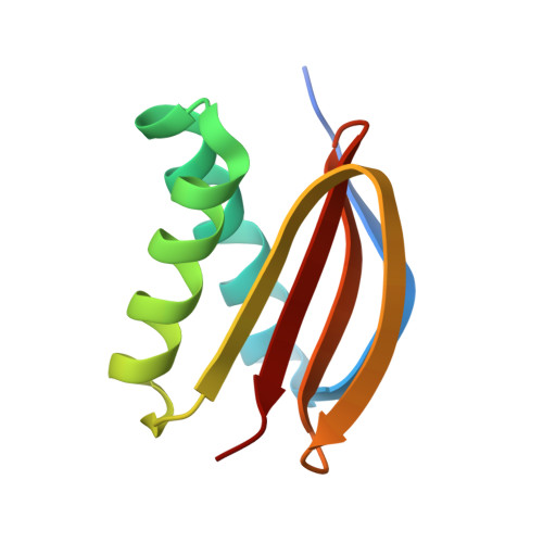
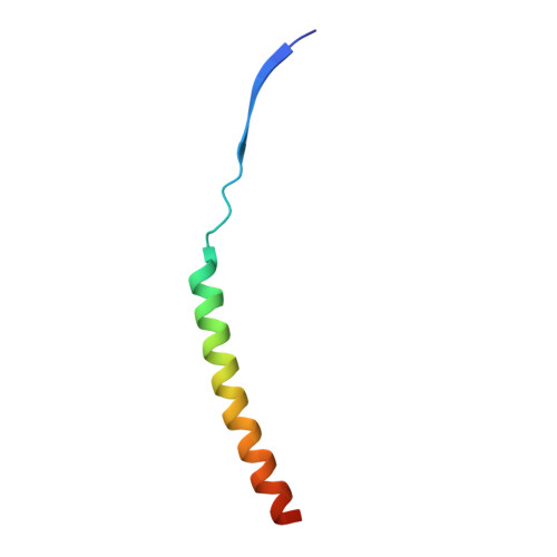
2XQQ, 3P8M - PubMed Abstract:
LC8 dynein light chain (DYNLL) is a eukaryotic hub protein that is thought to function as a dimerization engine. Its interacting partners are involved in a wide range of cellular functions. In its dozens of hitherto identified binding partners DYNLL binds to a linear peptide segment. The known segments define a loosely characterized binding motif: [D/S](-4)K(-3)X(-2)[T/V/I](-1)Q(0)[T/V](1)[D/E](2). The motifs are localized in disordered segments of the DYNLL-binding proteins and are often flanked by coiled coil or other potential dimerization domains. Based on a directed evolution approach, here we provide the first quantitative characterization of the binding preference of the DYNLL binding site. We displayed on M13 phage a naïve peptide library with seven fully randomized positions around a fixed, naturally conserved glutamine. The peptides were presented in a bivalent manner fused to a leucine zipper mimicking the natural dimer to dimer binding stoichiometry of DYNLL-partner complexes. The phage-selected consensus sequence V(-5)S(-4)R(-3)G(-2)T(-1)Q(0)T(1)E(2) resembles the natural one, but is extended by an additional N-terminal valine, which increases the affinity of the monomeric peptide twentyfold. Leu-zipper dimerization increases the affinity into the subnanomolar range. By comparing crystal structures of an SRGTQTE-DYNLL and a dimeric VSRGTQTE-DYNLL complex we find that the affinity enhancing valine is accommodated in a binding pocket on DYNLL. Based on the in vitro evolved sequence pattern we predict a large number of novel DYNLL binding partners in the human proteome. Among these EML3, a microtubule-binding protein involved in mitosis contains an exact match of the phage-evolved consensus and binds to DYNLL with nanomolar affinity. These results significantly widen the scope of the human interactome around DYNLL and will certainly shed more light on the biological functions and organizing role of DYNLL in the human and other eukaryotic interactomes.
Organizational Affiliation:
Department of Biochemistry, Eötvös Loránd University, Budapest, Hungary.