


Crystal Structure of peptidyl-tRNA hydrolase from Mycobacterium smegmatis at 2.2 A resolution
Kumar, A., Singh, A., Yadav, R., Sinha, M., Arora, A., Sharma, S., Singh, T.P.To be published.
Experimental Data Snapshot
wwPDB Validation 3D Report Full Report

Entity ID: 1 | |||||
|---|---|---|---|---|---|
| Molecule | Chains | Sequence Length | Organism | Details | Image |
| Peptidyl-tRNA hydrolase | 191 | Mycolicibacterium smegmatis MC2 155 | Mutation(s): 0 EC: 3.1.1.29 | 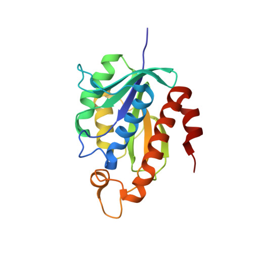 | |
UniProt | |||||
Find proteins for A0R3D3 (Mycolicibacterium smegmatis (strain ATCC 700084 / mc(2)155)) Explore A0R3D3 Go to UniProtKB: A0R3D3 | |||||
Entity Groups | |||||
| Sequence Clusters | 30% Identity50% Identity70% Identity90% Identity95% Identity100% Identity | ||||
| UniProt Group | A0R3D3 | ||||
Sequence AnnotationsExpand | |||||
| |||||
| Length ( Å ) | Angle ( ˚ ) |
|---|---|
| a = 44.909 | α = 90 |
| b = 58.943 | β = 90 |
| c = 61.854 | γ = 90 |
| Software Name | Purpose |
|---|---|
| DENZO | data reduction |
| AMoRE | phasing |
| REFMAC | refinement |
| SCALEPACK | data scaling |
RCSB PDB (citation) is hosted by
RCSB PDB is a member of the

