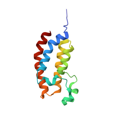
Crystal structure of the bromodomain of human CREBBP in complex with a hydroquinazolin ligand
Filippakopoulos, P., Picaud, S., Fedorov, O., Muniz, J., von Delft, F., Arrowsmith, C.H., Edwards, A.M., Weigelt, J., Bountra, C., Knapp, S., Structural Genomics Consortium (SGC)To be published.