


Crystal Structure of Acyl-CoA Dehydrogenase complexed with FAD from Bacillus anthracis
Kim, Y., Maltseva, N., Kwon, K., Anderson, W.F., Joachimiak, A., Center for Structural Genomics of Infectious Diseases (CSGID)To be published.
Experimental Data Snapshot
Entity ID: 1 | |||||
|---|---|---|---|---|---|
| Molecule | Chains | Sequence Length | Organism | Details | Image |
| Acyl-CoA dehydrogenase | 597 | Bacillus anthracis str. 'Ames Ancestor | Mutation(s): 0 Gene Names: BAS4875, BA_5246, GBAA5246, GBAA_5246 EC: 1.3.99 | 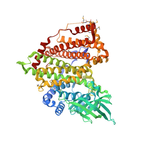 | |
UniProt | |||||
Find proteins for A0A6H3ADM7 (Bacillus anthracis) Explore A0A6H3ADM7 Go to UniProtKB: A0A6H3ADM7 | |||||
Entity Groups | |||||
| Sequence Clusters | 30% Identity50% Identity70% Identity90% Identity95% Identity100% Identity | ||||
| UniProt Group | A0A6H3ADM7 | ||||
Sequence AnnotationsExpand | |||||
| |||||
| Ligands 5 Unique | |||||
|---|---|---|---|---|---|
| ID | Chains | Name / Formula / InChI Key | 2D Diagram | 3D Interactions | |
| FAD Query on FAD | E [auth A], J [auth B], R [auth C], Y [auth D] | FLAVIN-ADENINE DINUCLEOTIDE C27 H33 N9 O15 P2 VWWQXMAJTJZDQX-UYBVJOGSSA-N |  | ||
| 1PE Query on 1PE | F [auth A], M [auth B], U [auth C], Z [auth D] | PENTAETHYLENE GLYCOL C10 H22 O6 JLFNLZLINWHATN-UHFFFAOYSA-N |  | ||
| PEG Query on PEG | G [auth A], N [auth B], W [auth C], X [auth D] | DI(HYDROXYETHYL)ETHER C4 H10 O3 MTHSVFCYNBDYFN-UHFFFAOYSA-N |  | ||
| SO4 Query on SO4 | I [auth A], O [auth B] | SULFATE ION O4 S QAOWNCQODCNURD-UHFFFAOYSA-L |  | ||
| GOL Query on GOL | H [auth A] K [auth B] L [auth B] P [auth B] Q [auth B] | GLYCEROL C3 H8 O3 PEDCQBHIVMGVHV-UHFFFAOYSA-N |  | ||
| Modified Residues 1 Unique | |||||
|---|---|---|---|---|---|
| ID | Chains | Type | Formula | 2D Diagram | Parent |
| MSE Query on MSE | A, B, C, D | L-PEPTIDE LINKING | C5 H11 N O2 Se |  | MET |
| Length ( Å ) | Angle ( ˚ ) |
|---|---|
| a = 75.613 | α = 92.8 |
| b = 98.028 | β = 106.63 |
| c = 107.743 | γ = 105.3 |
| Software Name | Purpose |
|---|---|
| REFMAC | refinement |
| HKL-3000 | data collection |
| BALBES | phasing |
| PHENIX | refinement |
| SBC-Collect | data collection |
| HKL-3000 | data reduction |
| HKL-3000 | data scaling |
RCSB PDB (citation) is hosted by
RCSB PDB is a member of the

