3OSG

The structure of protozoan parasite Trichomonas vaginalis Myb2 in complex with MRE-1-12 DNA


Experimental Data Snapshot

  • Method: X-RAY DIFFRACTION
  • Resolution: 2.00 Å
  • R-Value Free: 0.233 
  • R-Value Work: 0.191 
  • R-Value Observed: 0.193 

wwPDB Validation   3D Report Full Report


This is version 1.2 of the entry. See complete history


Literature

Molecular basis of the recognition of the ap65-1 gene transcription promoter elements by a Myb protein from the protozoan parasite Trichomonas vaginalis.

Jiang, I.Tsai, C.K.Chen, S.C.Wang, S.H.Amiraslanov, I.Chang, C.F.Wu, W.J.Tai, J.H.Liaw, Y.C.Huang, T.H.

(2011) Nucleic Acids Res 39: 8992-9008

  • DOI: https://doi.org/10.1093/nar/gkr558
  • Primary Citation of Related Structures:  
    3OSF, 3OSG

  • PubMed Abstract: 

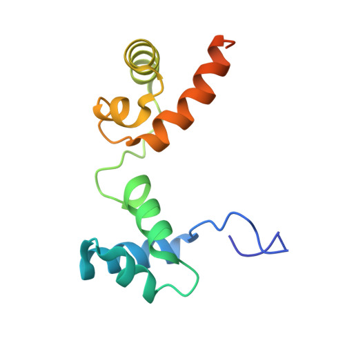
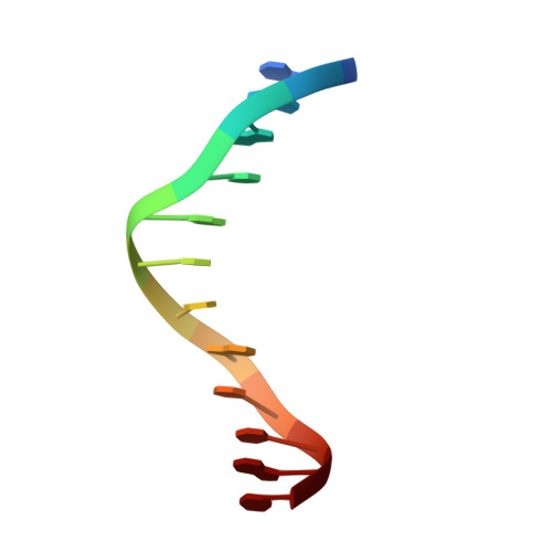
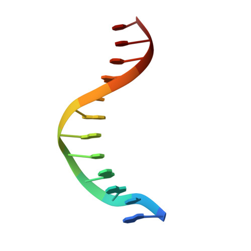
    Iron-inducible transcription of the ap65-1 gene in Trichomonas vaginalis involves at least three Myb-like transcriptional factors (tvMyb1, tvMyb2 and tvMyb3) that differentially bind to two closely spaced promoter sites, MRE-1/MRE-2r and MRE-2f. Here, we defined a fragment of tvMyb2 comprising residues 40-156 (tvMyb2₄₀₋₁₅₆) as the minimum structural unit that retains near full binding affinity with the promoter DNAs. Like c-Myb in vertebrates, the DNA-free tvMyb2₄₀₋₁₅₆ has a flexible and open conformation. Upon binding to the promoter DNA elements, tvMyb2₄₀₋₁₅₆ undergoes significant conformational re-arrangement and structure stabilization. Crystal structures of tvMyb2₄₀₋₁₅₆ in complex with promoter element-containing DNA oligomers showed that 5'-a/gACGAT-3' is the specific base sequence recognized by tvMyb2₄₀₋₁₅₆, which does not fully conform to that of the Myb binding site sequence. Furthermore, Lys⁴⁹, which is upstream of the R2 motif (amino acids 52-102) also participates in specific DNA sequence recognition. Intriguingly, tvMyb2₄₀₋₁₅₆ binds to the promoter elements in an orientation opposite to that proposed in the HADDOCK model of the tvMyb1₃₅₋₁₄₁/MRE-1-MRE-2r complex. These results shed new light on understanding the molecular mechanism of Myb-DNA recognition and provide a framework to study the molecular basis of transcriptional regulation of myriad Mybs in T. vaginalis.


  • Organizational Affiliation

    Institute of Biomedical Sciences, Academia Sinica, Taipei 115, Taiwan, ROC.


Macromolecules

Find similar proteins by:  (by identity cutoff)  |  3D Structure
Entity ID: 1
MoleculeChains Sequence LengthOrganismDetailsImage
MYB21
A, D
126Trichomonas vaginalisMutation(s): 0 
Gene Names: TVAG_211210
UniProt
Find proteins for Q58HP3 (Trichomonas vaginalis)
Explore Q58HP3 
Go to UniProtKB:  Q58HP3
Entity Groups  
Sequence Clusters30% Identity50% Identity70% Identity90% Identity95% Identity100% Identity
UniProt GroupQ58HP3
Sequence Annotations
Expand
  • Reference Sequence

Find similar nucleic acids by:  Sequence   |   3D Structure  

Entity ID: 2
MoleculeChains LengthOrganismImage
5'-D(*AP*AP*AP*TP*AP*TP*CP*GP*TP*TP*AP*T)-3'
B, E
12N/A
Sequence Annotations
Expand
  • Reference Sequence

Find similar nucleic acids by:  Sequence   |   3D Structure  

Entity ID: 3
MoleculeChains LengthOrganismImage
5'-D(*AP*TP*AP*AP*CP*GP*AP*TP*AP*TP*TP*T)-3'
C, F
12N/A
Sequence Annotations
Expand
  • Reference Sequence
Experimental Data & Validation

Experimental Data

  • Method: X-RAY DIFFRACTION
  • Resolution: 2.00 Å
  • R-Value Free: 0.233 
  • R-Value Work: 0.191 
  • R-Value Observed: 0.193 
  • Space Group: P 1 21 1
Unit Cell:
Length ( Å )Angle ( ˚ )
a = 40.115α = 90
b = 127.532β = 100.23
c = 41.473γ = 90
Software Package:
Software NamePurpose
HKL-2000data collection
MOLREPphasing
PHENIXrefinement
HKL-2000data reduction
HKL-2000data scaling

Structure Validation

View Full Validation Report


Entry History 

Deposition Data

Revision History  (Full details and data files)

  • Version 1.0: 2011-08-03
    Type: Initial release
  • Version 1.1: 2013-08-28
    Changes: Database references
  • Version 1.2: 2024-03-20
    Changes: Data collection, Database references