3OSE

Structure of the Kinase Associated Domain 1 (KA1) from MARK1 kinase


Experimental Data Snapshot

  • Method: X-RAY DIFFRACTION
  • Resolution: 1.70 Å
  • R-Value Free: 0.195 
  • R-Value Work: 0.175 
  • R-Value Observed: 0.176 

wwPDB Validation   3D Report Full Report


This is version 1.2 of the entry. See complete history


Literature

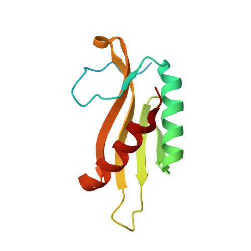
Kinase Associated-1 Domains Drive MARK/PAR1 Kinases to Membrane Targets by Binding Acidic Phospholipids.

Moravcevic, K.Mendrola, J.M.Schmitz, K.R.Wang, Y.H.Slochower, D.Janmey, P.A.Lemmon, M.A.

(2010) Cell 143: 966-977

  • DOI: https://doi.org/10.1016/j.cell.2010.11.028
  • Primary Citation of Related Structures:  
    3OSE, 3OSM, 3OST

  • PubMed Abstract: 

    Phospholipid-binding modules such as PH, C1, and C2 domains play crucial roles in location-dependent regulation of many protein kinases. Here, we identify the KA1 domain (kinase associated-1 domain), found at the C terminus of yeast septin-associated kinases (Kcc4p, Gin4p, and Hsl1p) and human MARK/PAR1 kinases, as a membrane association domain that binds acidic phospholipids. Membrane localization of isolated KA1 domains depends on phosphatidylserine. Using X-ray crystallography, we identified a structurally conserved binding site for anionic phospholipids in KA1 domains from Kcc4p and MARK1. Mutating this site impairs membrane association of both KA1 domains and intact proteins and reveals the importance of phosphatidylserine for bud neck localization of yeast Kcc4p. Our data suggest that KA1 domains contribute to "coincidence detection," allowing kinases to bind other regulators (such as septins) only at the membrane surface. These findings have important implications for understanding MARK/PAR1 kinases, which are implicated in Alzheimer's disease, cancer, and autism.


  • Organizational Affiliation

    Department of Biochemistry and Biophysics, University of Pennsylvania School of Medicine, Philadelphia, 19104, USA.


Macromolecules
Find similar proteins by:  (by identity cutoff)  |  3D Structure
Entity ID: 1
MoleculeChains Sequence LengthOrganismDetailsImage
Serine/threonine-protein kinase MARK1120Homo sapiensMutation(s): 0 
Gene Names: MARK1KIAA1477MARK
EC: 2.7.11.1
UniProt & NIH Common Fund Data Resources
Find proteins for Q9P0L2 (Homo sapiens)
Explore Q9P0L2 
Go to UniProtKB:  Q9P0L2
PHAROS:  Q9P0L2
GTEx:  ENSG00000116141 
Entity Groups  
Sequence Clusters30% Identity50% Identity70% Identity90% Identity95% Identity100% Identity
UniProt GroupQ9P0L2
Sequence Annotations
Expand
  • Reference Sequence
Experimental Data & Validation

Experimental Data

  • Method: X-RAY DIFFRACTION
  • Resolution: 1.70 Å
  • R-Value Free: 0.195 
  • R-Value Work: 0.175 
  • R-Value Observed: 0.176 
  • Space Group: P 31 2 1
Unit Cell:
Length ( Å )Angle ( ˚ )
a = 69.843α = 90
b = 69.843β = 90
c = 54.446γ = 120
Software Package:
Software NamePurpose
HKL-2000data collection
PHASERphasing
REFMACrefinement
HKL-2000data reduction
HKL-2000data scaling

Structure Validation

View Full Validation Report


Entry History 

Deposition Data

Revision History  (Full details and data files)

  • Version 1.0: 2010-12-22
    Type: Initial release
  • Version 1.1: 2011-07-13
    Changes: Version format compliance
  • Version 1.2: 2024-02-21
    Changes: Data collection, Database references, Derived calculations