3ORG

Crystal Structure of a eukaryotic CLC transporter


Experimental Data Snapshot

  • Method: X-RAY DIFFRACTION
  • Resolution: 3.50 Å
  • R-Value Free: 0.284 
  • R-Value Work: 0.259 
  • R-Value Observed: 0.261 

wwPDB Validation   3D Report Full Report


This is version 1.2 of the entry. See complete history


Literature

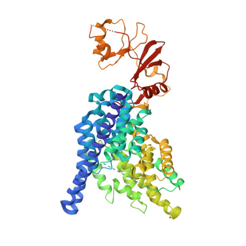
Structure of a eukaryotic CLC transporter defines an intermediate state in the transport cycle.

Feng, L.Campbell, E.B.Hsiung, Y.MacKinnon, R.

(2010) Science 330: 635-641

  • DOI: https://doi.org/10.1126/science.1195230
  • Primary Citation of Related Structures:  
    3ORG

  • PubMed Abstract: 

    CLC proteins transport chloride (Cl(-)) ions across cell membranes to control the electrical potential of muscle cells, transfer electrolytes across epithelia, and control the pH and electrolyte composition of intracellular organelles. Some members of this protein family are Cl(-) ion channels, whereas others are secondary active transporters that exchange Cl(-) ions and protons (H(+)) with a 2:1 stoichiometry. We have determined the structure of a eukaryotic CLC transporter at 3.5 angstrom resolution. Cytoplasmic cystathionine beta-synthase (CBS) domains are strategically positioned to regulate the ion-transport pathway, and many disease-causing mutations in human CLCs reside on the CBS-transmembrane interface. Comparison with prokaryotic CLC shows that a gating glutamate residue changes conformation and suggests a basis for 2:1 Cl(-)/H(+) exchange and a simple mechanistic connection between CLC channels and transporters.


  • Organizational Affiliation

    Laboratory of Molecular Neurobiology and Biophysics, Rockefeller University, Howard Hughes Medical Institute, 1230 York Avenue, New York, NY 10065, USA.


Macromolecules
Find similar proteins by:  (by identity cutoff)  |  3D Structure
Entity ID: 1
MoleculeChains Sequence LengthOrganismDetailsImage
CmCLC
A, B, C, D
632Cyanidioschyzon merolaeMutation(s): 0 
Gene Names: CLC
Entity Groups  
Sequence Clusters30% Identity50% Identity70% Identity90% Identity95% Identity100% Identity
Sequence Annotations
Expand
  • Reference Sequence
Experimental Data & Validation

Experimental Data

  • Method: X-RAY DIFFRACTION
  • Resolution: 3.50 Å
  • R-Value Free: 0.284 
  • R-Value Work: 0.259 
  • R-Value Observed: 0.261 
  • Space Group: C 1 2 1
Unit Cell:
Length ( Å )Angle ( ˚ )
a = 229.629α = 90
b = 178.27β = 129.21
c = 145.135γ = 90
Software Package:
Software NamePurpose
DENZOdata reduction
SCALEPACKdata scaling
REFMACrefinement
PDB_EXTRACTdata extraction
HKL-2000data collection
HKL-2000data reduction
HKL-2000data scaling
SHELXSphasing

Structure Validation

View Full Validation Report


Entry History 

Deposition Data

Revision History  (Full details and data files)

  • Version 1.0: 2010-10-06
    Type: Initial release
  • Version 1.1: 2011-07-13
    Changes: Version format compliance
  • Version 1.2: 2024-02-21
    Changes: Data collection, Database references, Derived calculations