3OQ6

Horse liver alcohol dehydrogenase A317C mutant complexed with NAD+ and 2,3,4,5,6-pentafluorobenzyl alcohol


Experimental Data Snapshot

  • Method: X-RAY DIFFRACTION
  • Resolution: 1.20 Å
  • R-Value Free: 0.162 
  • R-Value Work: 0.135 
  • R-Value Observed: 0.135 

wwPDB Validation   3D Report Full Report


Ligand Structure Quality Assessment 


This is version 1.2 of the entry. See complete history


Literature

Origins of the high catalytic activity of human alcohol dehydrogenase 4 studied with horse liver A317C alcohol dehydrogenase.

Herdendorf, T.J.Plapp, B.V.

(2011) Chem Biol Interact 191: 42-47

  • DOI: https://doi.org/10.1016/j.cbi.2010.12.015
  • Primary Citation of Related Structures:  
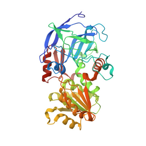
    3OQ6

  • PubMed Abstract: 

    The turnover numbers and other kinetic constants for human alcohol dehydrogenase (ADH) 4 ("stomach" isoenzyme) are substantially larger (10-100-fold) than those for human class I and horse liver alcohol dehydrogenases. Comparison of the primary amino acid sequences (69% identity) and tertiary structures of these enzymes led to the suggestion that residue 317, which makes a hydrogen bond with the nicotinamide amide nitrogen of the coenzyme, may account for these differences. Ala-317 in the class I enzymes is substituted with Cys in human ADH4, and locally different conformations of the peptide backbones could affect coenzyme binding. This hypothesis was tested by making the A317C substitution in horse liver ADH1E and comparisons to the wild-type ADH1E. The steady-state kinetic constants for the oxidation of benzyl alcohol and the reduction of benzaldehyde catalyzed by the A317C enzyme were very similar (up to about 2-fold differences) to those for the wild-type enzyme. Transient kinetics showed that the rate constants for binding of NAD(+) and NADH were also similar. Transient reaction data were fitted to the full Ordered Bi Bi mechanism and showed that the rate constants for hydride transfer decreased by about 2.8-fold with the A317C substitution. The structure of A317C ADH1E complexed with NAD(+) and 2,3,4,5,6-pentafluorobenzyl alcohol at 1.2 Å resolution is essentially identical to the structure of the wild-type enzyme, except near residue 317 where the additional sulfhydryl group displaces a water molecule that is present in the wild-type enzyme. ADH is adaptable and can tolerate internal substitutions, but the protein dynamics apparently are affected, as reflected in rates of hydride transfer. The A317C substitution is not solely responsible for the larger kinetic constants in human ADH4; thus, the differences in catalytic activity must arise from one or more of the other hundred substitutions in the enzyme.


  • Organizational Affiliation

    Department of Biochemistry, The University of Iowa, Iowa City, IA 52242-1109, USA.


Macromolecules
Find similar proteins by:  (by identity cutoff)  |  3D Structure
Entity ID: 1
MoleculeChains Sequence LengthOrganismDetailsImage
Alcohol dehydrogenase E chain
A, B
374Equus caballusMutation(s): 1 
Gene Names: ADH E
EC: 1.1.1.1
UniProt
Find proteins for P00327 (Equus caballus)
Explore P00327 
Go to UniProtKB:  P00327
Entity Groups  
Sequence Clusters30% Identity50% Identity70% Identity90% Identity95% Identity100% Identity
UniProt GroupP00327
Sequence Annotations
Expand
  • Reference Sequence
Small Molecules
Ligands 4 Unique
IDChains Name / Formula / InChI Key2D Diagram3D Interactions
NAJ
Query on NAJ

Download Ideal Coordinates CCD File 
E [auth A],
K [auth B]
NICOTINAMIDE-ADENINE-DINUCLEOTIDE (ACIDIC FORM)
C21 H27 N7 O14 P2
BAWFJGJZGIEFAR-NNYOXOHSSA-N
PFB
Query on PFB

Download Ideal Coordinates CCD File 
F [auth A],
L [auth B]
2,3,4,5,6-PENTAFLUOROBENZYL ALCOHOL
C7 H3 F5 O
PGJYYCIOYBZTPU-UHFFFAOYSA-N
MRD
Query on MRD

Download Ideal Coordinates CCD File 
G [auth A],
H [auth A],
M [auth B]
(4R)-2-METHYLPENTANE-2,4-DIOL
C6 H14 O2
SVTBMSDMJJWYQN-RXMQYKEDSA-N
ZN
Query on ZN

Download Ideal Coordinates CCD File 
C [auth A],
D [auth A],
I [auth B],
J [auth B]
ZINC ION
Zn
PTFCDOFLOPIGGS-UHFFFAOYSA-N
Experimental Data & Validation

Experimental Data

  • Method: X-RAY DIFFRACTION
  • Resolution: 1.20 Å
  • R-Value Free: 0.162 
  • R-Value Work: 0.135 
  • R-Value Observed: 0.135 
  • Space Group: P 1
Unit Cell:
Length ( Å )Angle ( ˚ )
a = 44.34α = 91.98
b = 51.31β = 102.95
c = 92.21γ = 110.11
Software Package:
Software NamePurpose
d*TREKdata scaling
REFMACrefinement
PDB_EXTRACTdata extraction
d*TREKdata reduction

Structure Validation

View Full Validation Report


Ligand Structure Quality Assessment 


Entry History 

Deposition Data

Revision History  (Full details and data files)

  • Version 1.0: 2010-11-10
    Type: Initial release
  • Version 1.1: 2011-07-13
    Changes: Version format compliance
  • Version 1.2: 2023-09-06
    Changes: Data collection, Database references, Derived calculations, Refinement description