


The structure of a proline dipeptidase from Streptococcus agalactiae 2603V
Fan, Y., Wu, R., Morales, J., Clancy, S., Joachimiak, A.To be published.
Experimental Data Snapshot
wwPDB Validation 3D Report Full Report

Entity ID: 1 | |||||
|---|---|---|---|---|---|
| Molecule | Chains | Sequence Length | Organism | Details | Image |
| Proline dipeptidase | 132 | Streptococcus agalactiae 2603V/R | Mutation(s): 0 Gene Names: pepQ, SAG0706 | 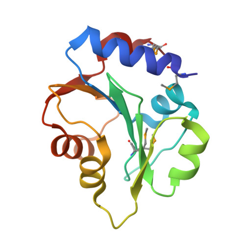 | |
UniProt | |||||
Find proteins for Q8E0M4 (Streptococcus agalactiae serotype V (strain ATCC BAA-611 / 2603 V/R)) Explore Q8E0M4 Go to UniProtKB: Q8E0M4 | |||||
Entity Groups | |||||
| Sequence Clusters | 30% Identity50% Identity70% Identity90% Identity95% Identity100% Identity | ||||
| UniProt Group | Q8E0M4 | ||||
Sequence AnnotationsExpand | |||||
| |||||
| Modified Residues 1 Unique | |||||
|---|---|---|---|---|---|
| ID | Chains | Type | Formula | 2D Diagram | Parent |
| MSE Query on MSE | A, B | L-PEPTIDE LINKING | C5 H11 N O2 Se |  | MET |
| Length ( Å ) | Angle ( ˚ ) |
|---|---|
| a = 43.744 | α = 90 |
| b = 58.325 | β = 90 |
| c = 101.995 | γ = 90 |
| Software Name | Purpose |
|---|---|
| SBC-Collect | data collection |
| SHELXD | phasing |
| MLPHARE | phasing |
| ARP | model building |
| WARP | model building |
| HKL-3000 | phasing |
| REFMAC | refinement |
| HKL-3000 | data reduction |
| HKL-3000 | data scaling |
RCSB PDB (citation) is hosted by
RCSB PDB is a member of the

