3ONV

Crystal structure of E. Coli purine nucleoside phosphorylase with SO4


Experimental Data Snapshot

  • Method: X-RAY DIFFRACTION
  • Resolution: 1.89 Å
  • R-Value Free: 0.203 
  • R-Value Work: 0.175 
  • R-Value Observed: 0.176 

wwPDB Validation   3D Report Full Report


This is version 1.3 of the entry. See complete history


Literature

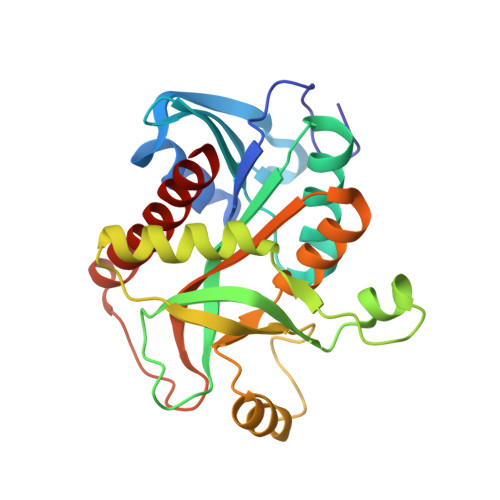
Validation of the catalytic mechanism of Escherichia coli purine nucleoside phosphorylase by structural and kinetic studies.

Mikleusevic, G.Stefanic, Z.Narczyk, M.Wielgus-Kutrowska, B.Bzowska, A.Luic, M.

(2011) Biochimie 93: 1610-1622

  • DOI: https://doi.org/10.1016/j.biochi.2011.05.030
  • Primary Citation of Related Structures:  
    3ONV, 3OOE, 3OOH, 3OPV

  • PubMed Abstract: 

    The catalytic mechanism of Escherichia coli purine nucleoside phosphorylase (PNP) is revised using site-directed mutagenesis, kinetic studies and structure determinations. The experimental evidence on the role of the particular catalytic amino acid during catalysis has not been available. Therefore, the active site mutants Arg24Ala, Asp204Ala, Asp204Asn, Arg217Ala and Asp204Ala/Arg217Ala were prepared and their kinetics and thermodynamic studies were carried out. The activity tests with natural substrates and 7-methylguanosine confirmed the earlier hypothesis, that catalysis involves protonation of the purine base at position N7 by Asp204, which is triggered by Arg217. The crystal structures of the wild type in complexes with phosphate and sulphate, respectively, and of the Arg24Ala mutant in complex with phosphate/sulphate were determined. The structural data show that previously observed conformational change is a result of the phosphate binding and its interaction with Arg24. As E. coli PNP is a promising candidate for the tumour-directed gene therapy, our results may also help to design efficient mutants useful in gene therapy.


  • Organizational Affiliation

    Division of Physical Chemistry, Ruđer Bošković Institute, POB 180, HR-10002 Zagreb, Croatia.


Macromolecules
Find similar proteins by:  (by identity cutoff)  |  3D Structure
Entity ID: 1
MoleculeChains Sequence LengthOrganismDetailsImage
Purine nucleoside phosphorylase deoD-type
A, B, C
237Escherichia coli K-12Mutation(s): 0 
Gene Names: deoDEcDH1_3614
EC: 2.4.2.1
UniProt
Find proteins for P0ABP8 (Escherichia coli (strain K12))
Explore P0ABP8 
Go to UniProtKB:  P0ABP8
Entity Groups  
Sequence Clusters30% Identity50% Identity70% Identity90% Identity95% Identity100% Identity
UniProt GroupP0ABP8
Sequence Annotations
Expand
  • Reference Sequence
Experimental Data & Validation

Experimental Data

  • Method: X-RAY DIFFRACTION
  • Resolution: 1.89 Å
  • R-Value Free: 0.203 
  • R-Value Work: 0.175 
  • R-Value Observed: 0.176 
  • Space Group: P 61 2 2
Unit Cell:
Length ( Å )Angle ( ˚ )
a = 120.304α = 90
b = 120.304β = 90
c = 239.809γ = 120
Software Package:
Software NamePurpose
PHENIXrefinement
XSCALEdata scaling
PDB_EXTRACTdata extraction
MAR345data collection
XDSdata reduction
PHENIXphasing

Structure Validation

View Full Validation Report


Entry History 

Deposition Data

Revision History  (Full details and data files)

  • Version 1.0: 2011-07-13
    Type: Initial release
  • Version 1.1: 2013-08-14
    Changes: Database references
  • Version 1.2: 2017-11-08
    Changes: Refinement description
  • Version 1.3: 2024-03-20
    Changes: Data collection, Database references, Derived calculations