3OIS

Crystal Structure Xylellain, a cysteine protease from Xylella fastidiosa


Experimental Data Snapshot

  • Method: X-RAY DIFFRACTION
  • Resolution: 1.65 Å
  • R-Value Free: 0.216 
  • R-Value Work: 0.166 
  • R-Value Observed: 0.169 

wwPDB Validation   3D Report Full Report


Ligand Structure Quality Assessment 


This is version 1.2 of the entry. See complete history


Literature

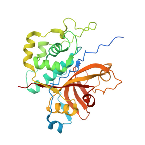
The crystal structure of the cysteine protease Xylellain from Xylella fastidiosa reveals an intriguing activation mechanism.

Leite, N.R.Faro, A.R.Dotta, M.A.Faim, L.M.Gianotti, A.Silva, F.H.Oliva, G.Thiemann, O.H.

(2013) FEBS Lett 587: 339-344

  • DOI: https://doi.org/10.1016/j.febslet.2013.01.009
  • Primary Citation of Related Structures:  
    3OIS

  • PubMed Abstract: 

    Xylella fastidiosa is responsible for a wide range of economically important plant diseases. We report here the crystal structure and kinetic data of Xylellain, the first cysteine protease characterized from the genome of the pathogenic X. fastidiosa strain 9a5c. Xylellain has a papain-family fold, and part of the N-terminal sequence blocks the enzyme active site, thereby mediating protein activity. One novel feature identified in the structure is the presence of a ribonucleotide bound outside the active site. We show that this ribonucleotide plays an important regulatory role in Xylellain enzyme kinetics, possibly functioning as a physiological mediator.


  • Organizational Affiliation

    Instituto de Física de São Carlos, Universidade de São Paulo, São Carlos, SP 13566-590, Brazil.


Macromolecules
Find similar proteins by:  (by identity cutoff)  |  3D Structure
Entity ID: 1
MoleculeChains Sequence LengthOrganismDetailsImage
Cysteine protease
A, B, C, D
291Xylella fastidiosaMutation(s): 0 
Gene Names: XF_0156
UniProt
Find proteins for Q9PGZ0 (Xylella fastidiosa (strain 9a5c))
Explore Q9PGZ0 
Go to UniProtKB:  Q9PGZ0
Entity Groups  
Sequence Clusters30% Identity50% Identity70% Identity90% Identity95% Identity100% Identity
UniProt GroupQ9PGZ0
Sequence Annotations
Expand
  • Reference Sequence
Experimental Data & Validation

Experimental Data

  • Method: X-RAY DIFFRACTION
  • Resolution: 1.65 Å
  • R-Value Free: 0.216 
  • R-Value Work: 0.166 
  • R-Value Observed: 0.169 
  • Space Group: P 1
Unit Cell:
Length ( Å )Angle ( ˚ )
a = 55.089α = 75.86
b = 69.314β = 75.43
c = 82.368γ = 66.51
Software Package:
Software NamePurpose
MOSFLMdata reduction
SCALAdata scaling
SOLVEphasing
RESOLVEphasing
REFMACrefinement
PDB_EXTRACTdata extraction
MAR345dtbdata collection

Structure Validation

View Full Validation Report


Ligand Structure Quality Assessment 


Entry History 

Deposition Data

Revision History  (Full details and data files)

  • Version 1.0: 2011-08-03
    Type: Initial release
  • Version 1.1: 2013-03-13
    Changes: Database references
  • Version 1.2: 2024-02-21
    Changes: Data collection, Database references, Derived calculations