3OFS

Dynamic conformations of the CD38-mediated NAD cyclization captured using multi-copy crystallography


Experimental Data Snapshot

  • Method: X-RAY DIFFRACTION
  • Resolution: 2.20 Å
  • R-Value Free: 0.250 
  • R-Value Work: 0.203 
  • R-Value Observed: 0.206 

wwPDB Validation   3D Report Full Report


Ligand Structure Quality Assessment 


This is version 1.3 of the entry. See complete history


Literature

Dynamic Conformations of the CD38-Mediated NAD Cyclization Captured in a Single Crystal

Zhang, H.Graeff, R.Chen, Z.Zhang, L.R.Zhang, L.H.Lee, H.C.Hao, Q.

(2011) J Mol Biol 405: 1070-1078

  • DOI: https://doi.org/10.1016/j.jmb.2010.11.044
  • Primary Citation of Related Structures:  
    3OFS

  • PubMed Abstract: 

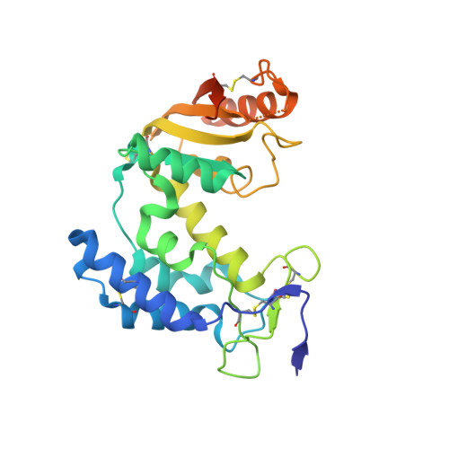
    The extracellular domain of human CD38 is a multifunctional enzyme involved in the metabolism of two Ca(2+) messengers: cyclic ADP-ribose and nicotinic acid adenine dinucleotide phosphate. When NAD is used as substrate, CD38 predominantly hydrolyzes it to ADP-ribose, with a trace amount of cyclic ADP-ribose produced through cyclization of the substrate. However, mutation of a key residue at the active site, E146, inhibits the hydrolysis activity of CD38 but greatly increases its cyclization activity. To understand the role of the residue E146 in the catalytic process, we determined the crystal structure of the E146A mutant protein with a substrate analogue, arabinosyl-2'-fluoro-deoxy-nicotinamide adenine dinucleotide. The structure captured the enzymatic reaction intermediates in six different conformations in a crystallographic asymmetric unit. The structural results indicate a folding-back process for the adenine ring of the substrate and provide the first multiple snapshots of the process. Our approach of utilizing multiple molecules in the crystallographic asymmetric unit should be generally applicable for capturing the dynamic nature of enzymatic catalysis.


  • Organizational Affiliation

    Department of Physiology, The University of Hong Kong, Hong Kong SAR, China.


Macromolecules
Find similar proteins by:  (by identity cutoff)  |  3D Structure
Entity ID: 1
MoleculeChains Sequence LengthOrganismDetailsImage
ADP-ribosyl cyclase 1
A, B, C, D, E
A, B, C, D, E, F
257Homo sapiensMutation(s): 6 
Gene Names: CD38
EC: 3.2.2.5
UniProt & NIH Common Fund Data Resources
Find proteins for P28907 (Homo sapiens)
Explore P28907 
Go to UniProtKB:  P28907
PHAROS:  P28907
GTEx:  ENSG00000004468 
Entity Groups  
Sequence Clusters30% Identity50% Identity70% Identity90% Identity95% Identity100% Identity
UniProt GroupP28907
Sequence Annotations
Expand
  • Reference Sequence
Experimental Data & Validation

Experimental Data

  • Method: X-RAY DIFFRACTION
  • Resolution: 2.20 Å
  • R-Value Free: 0.250 
  • R-Value Work: 0.203 
  • R-Value Observed: 0.206 
  • Space Group: P 21 21 21
Unit Cell:
Length ( Å )Angle ( ˚ )
a = 56.655α = 90
b = 146.662β = 90
c = 191.086γ = 90
Software Package:
Software NamePurpose
DENZOdata reduction
SCALEPACKdata scaling
REFMACrefinement
PDB_EXTRACTdata extraction

Structure Validation

View Full Validation Report


Ligand Structure Quality Assessment 


Entry History 

Deposition Data

Revision History  (Full details and data files)

  • Version 1.0: 2010-12-15
    Type: Initial release
  • Version 1.1: 2011-07-13
    Changes: Version format compliance
  • Version 1.2: 2017-11-08
    Changes: Refinement description
  • Version 1.3: 2023-11-01
    Changes: Data collection, Database references, Derived calculations, Refinement description