Chemically Self-Assembled Antibody Nanorings (CSANs): Design and Characterization of an Anti-CD3 IgM Biomimetic.
Li, Q., So, C.R., Fegan, A., Cody, V., Sarikaya, M., Vallera, D.A., Wagner, C.R.(2010) J Am Chem Soc 132: 17247-17257
- PubMed: 21077608
- DOI: https://doi.org/10.1021/ja107153a
- Primary Citation of Related Structures:
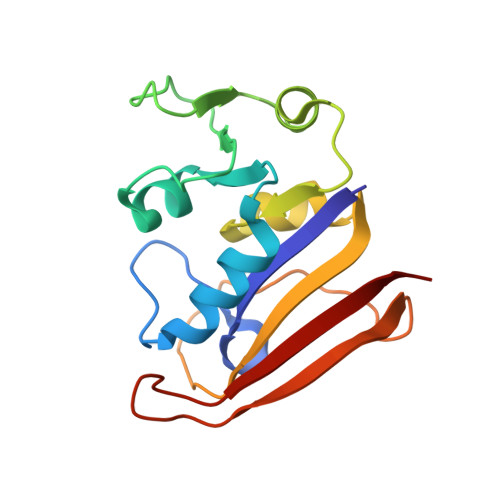
3OCH - PubMed Abstract:
A number of clever recombinant methodologies have been developed that recapitulate the valencies of IgG's (bivalent) and IgA's (tetravalent). Although higher synthetic valencies have been achieved by conjugation of either monoclonal antibodies or single-chain antibodies to nanoparticles and liposomes, a method for the preparation of recombinant antibodies with valencies similar to IgM's (decavalent) but considerably less than what is generally found after antibody particle conjugation has yet to be devised. Recently, we have developed a methodology for the design of bivalent Chemically Self-Assembled Antibody Nanorings (CSANs). We now report the crystal structure of the nanoring subunit composed of the E. coli DHFR dimer and a methotrexate dimerizer (MTX2-C9) containing a visible nine methylene linker and a protocol for the preparation of CSANs from this subunit with valencies similar to IgM's, ranging from 8-10 single chain antibodies (scFvs). The multivalent CSANs were reversibly assembled from a fusion protein dihydrofolate reductase (DHFR)-DHFR-antiCD3 scFv containing a single glycine linker between the two DHFR scaffolding proteins. We also demonstrate that, similar to the parental bivalent anti-CD3 monoclonal antibody (mAB), anti-CD3 CSANs selectively bind to CD3+ leukemia cells and undergo rapid internalization through a caveolin-independent pathway that requires cholesterol, actin polymerization, and protein tyrosine kinase activation. While treatment with the monoclonal antibody leads to T-cell activation and nearly complete loss (i.e., 90%) of the surface displayed T-cell receptor (TCR), only 25-30% of the TCR down regulate and no significant T-cell proliferation is observed after treatment of peripheral blood mononuclear cells (PBMCs) with anti-CD3 CSANs. Consistent with the proliferation findings, 15-25% less CD25 (IL-2 receptor) was found on the surface of PBMCs treated with either the polyvalent or bivalent anti-CD3 CSANs, respectively, than on PBMCs treated with the parental mAB. Comparative experiments with F(ab')2 derived from the mAB confirm that the activation of the T-cells by the mAB is dependent on the Fc domain, and thus interactions of the PBMC T-cells with accessory cells, such as macrophages. Taken together, our results demonstrate that anti-CD3 CSANs with valencies ranging from 2 to 8 could be employed for radionuclide, drug, or potentially oligonucleotide delivery to T-cells without, as has been observed for other antibody conjugated nanoparticles, the deleterious effects of activation observed for mAB. Further the CSAN construct may be adapted for the preparation of other multivalent scFvs.
Organizational Affiliation:
Department of Chemistry, University of Minnesota, Minneapolis, Minnesota 55455, USA.